#diagnosticimaging
Diagnostic Imaging Rotation
I had been dreading this rotation all year and it was finally upon me. Countless stories of students crying and having breakdowns were circulating and didn’t inspire a great deal of enthusiasm. The first couple of days were really quite overwhelming. It seemed as though there was a new assessment every five minutes!
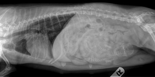
Each day generally began with an online quiz for which we frantically revised the night before. Once finished, we would convene in front of the light boxes for interpretation rounds. This involved five students being selected at random to look at a series of radiographs we’d never seen before, interpret them using a systematic approach, and arrive at a diagnosis. This was both timed and assessed. Each day would cover a different topic: musculoskeletal, thorax, abdomen, equine, and so on. In a desperate attempt to beat the tears, our group diffused the tension with humour. I don’t remember how it started, but we began referring to interpretation rounds as ‘the grilling’ or ‘the roasting’. The puns were endless. When asked if we were ready, the response was “I’ve been marinating all night!”. As the first person of the day stepped up to the hot seat, someone would mime lighting a grill. If the radiologist was being especially harsh, we’d say “the grill is hot today!”, and if they were giving someone a hard time, someone would make a sizzling noise in the background. Laughter was the only way to get through it.
On the first day, we had a short ultrasonography tutorial and a quick practice scanning a nurse’s dog. Each student was assigned a day of ultrasonography during which we observed the specialist performing scans and pretended to understand the grey shapes on the screen. Another day was spent on ‘interpretations’ where we interpreted all of the radiographs taken that day. The remaining students were assigned to radiography, which involved positioning real patients and taking the radiographs according to requests from clinicians. These were also timed and assessed, with many ways to instantly fail. If there weren’t any real patients, we were assessed on the dummy dog (named Emily) who was frustratingly inflexible. In the evenings, we would convene again for rounds and share any interesting cases with the group.
Towards the end of the second week, we had yet another timed exam where we had to take radiographs of a horse’s foot, fetlock or carpus. I forgot to check the exposure parameters on one of my shots, but it happened to be on the right setting by coincidence. On the final morning, we had an online theory exam.
As much as it pains me to admit it, I think I actually enjoyed diagnostic imaging! It was great to finally learn how to read radiographs and I improved considerably over the two weeks. Not a single tear was shed in our group, which was perhaps our greatest achievement.
Post link

