#eye movements

When something gets in the way of our ability to see, we quickly pick up a new way to look, in much the same way that we would learn to ride a bike, according to a new study published in the Cell Press journal Current Biology on August 15.
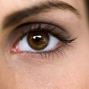
Our eyes are constantly on the move, darting this way and that four to five times per second. Now researchers have found that the precise manner of those eye movements can change within a matter of hours. This discovery by researchers from the University of Southern California might suggest a way to help those with macular degeneration better cope with vision loss.
“The system that controls how the eyes move is far more malleable than the literature has suggested,” says Bosco Tjan of the University of Southern California. “We showed that people with normal vision can quickly adjust to a temporary occlusion of their foveal vision by adapting a consistent point in their peripheral vision as their new point of gaze.”
The fovea refers to the small, center-most portion of the retina, which is responsible for our high-resolution vision. We move our eyes to direct the fovea to different parts of a scene, constructing a picture of the world around us. In those with age-related macular degeneration, progressive loss of foveal vision leads to visual impairment and blindness.
In the new study, MiYoung Kwon, Anirvan Nandy, and Tjan simulated a loss of foveal vision in six normally sighted young adults by blocking part of a visual scene with a gray disc that followed the individuals’ eye gaze. Those individuals were then asked to complete demanding object-following and visual-search tasks. Within three hours of working on those tasks, people showed a remarkably fast and spontaneous adjustment of eye movements. Once developed, that change in their “point of gaze” was retained over a period of weeks and was reengaged whenever their foveal vision was blocked.
Tjan and his team say they were surprised by the rate of this adjustment. They note that patients with macular degeneration frequently do adapt their point of gaze, but in a process that takes months, not days or hours. They suggest that practice with a visible gray disc like the one used in the study might help speed that process of visual rehabilitation along. The discovery also reveals that the oculomotor (eye movement) system prefers control simplicity over optimality.
“Gaze control by the oculomotor system, although highly automatic, is malleable in the same sense that motor control of the limbs is malleable,” Tjan says. “This finding is potentially very good news for people who lose their foveal vision due to macular diseases. It may be possible to create the right conditions for the oculomotor system to quickly adjust,” Kwon adds.
Amyotrophic lateral sclerosis (ALS) is a lethal disease characterised by progressive loss of motor neurons and subsequent muscle atrophy, weakness and paralysis. However, certain motor neuron groups are, for unknown reasons, relatively resistant to degeneration in ALS. Among the most resistant are oculomotor neurons, which are located in the brain stem and control eye movement. Consequently, eye-tracking devices are used to enable paralyzed ALS patients to communicate through computers.
Incurable disease
The disease is currently incurable, but researchers are looking for genes that can be used to develop treatments able to arrest the progress of the disease and prevent the loss of motor neurons. To this end, Eva Hedlund’s team at Karolinska Institutet’s Department of Neuroscience has focused its attention on the oculomotor neurons.
“We have now identified a factor, insulin-like growth factor 2, or IGF-2, within the resistant oculomotor neurons. We show that IGF-2 can rescue human motor neurons from degenerating,” explains Dr Hedlund, who has led the study with Stefania Corti at Milano University.
The researchers used skin cells obtained from ALS patients. The skin cells were reprogrammed into stem cells and further developed into motor neurons. Upon receiving IGF-2 the neurons fared better than normally under conditions harmful to motor neurons.
Positive effect
Since IGF-2 had a positive effect on cultivated neurons outside the body, the researchers proceeded to examine if it could also protect motor neurons in mice that develop ALS-like disease. When the researcher gave the ALS mice IGF-2 through gene therapy they lived longer.
“We can see that motor neurons are preserved and that IGF-2 treatment causes the axons to regenerate and recreate vital connections with muscles that were previously lost,” says Dr Hedlund.
The closely related hormone IGF-1 has been clinically tested through administration under the skin to patients with ALS, but the results have been contradictory. So far IGF-2 has not been tested in a clinical setting. The new study supports the idea that administration of IGF-2 or IGF-1 directly to motor neurons through gene therapy could have a positive effect.