#orbitofrontal cortex

How to make up your mind when the glass seems half empty?
Is a new high-income job offer worth accepting if it means commuting an extra hour to work? People often have to make tough choices regarding whether to endure some level of discomfort to take advantage of an opportunity or otherwise walk away from the reward. In making such choices, it turns out that the brain weighs our desire to go for the reward against our desire to avoid the related hardship.
In previous research, negative mental states have been shown to upset this balance between payoff and hardship toward more ‘pessimistic’ decision making and avoidance. For example, scientists know that people experiencing anxiety have a stronger-than-normal desire to avoid negative consequences. And people with depression have a weaker desire to approach the reward in the first place. But there is still much we do not know about how the brain incorporates feelings into decision making.
Neuroscientists at Kyoto University’s Institute for Advanced Study of Human Biology (WPI-ASHBi) have connected some of the dots to reveal the brain networks that give anxiety influence over decisions. Writing in the journal Frontiers in Neuroscience, the group has published a review that synthesizes results from years of brain measurements in rats and primates and relates these findings to the human brain.
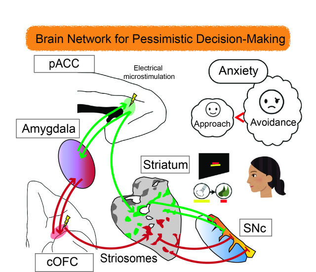
“We are facing a new epidemic of anxiety, and it is important that we understand how our anxiety influences our decision making,” says Ken-ichi Amemori, associate professor in neuroscience at Kyoto University, ASHBi. “There is a real need for a better understanding of what is happening in the brain here. It is very difficult for us to see exactly where and how anxiety manifests in humans, but studies in primate brains have pointed to neurons in the ACC [anterior cingulate cortex] as being important in these decision-making processes.”
Thinking of the brain as an onion, the ACC lies in a middle layer, wrapping around the tough ‘heart’, or corpus callosum, which joins the two hemispheres. The ACC is also well-connected with many other parts of the brain controlling higher and lower functions with a role in integrating feelings with rational thinking.
The team started by measuring brain activity in rhesus macaques while they performed a task to select or reject a reward in the form of food combined with different levels of ‘punishment’ in the form of an annoying blast of air in the face. The potential choices were visually represented on a screen, and the monkeys used a joystick to make their selection, revealing how much discomfort they were willing to consider acceptable.
When the team probed the ACC of the monkeys, they identified groups of neurons that activated or deactivated in line with the sizes of the reward or punishment on offer. The neurons associated with avoidance and pessimistic decision-making were particularly concentrated in a part of the ACC called the pregenual ACC (pACC). This region has been previously linked to major depressive disorder and generalized anxiety disorder in humans.
Microstimulation of the pACC with a low-level electrical pulse caused the monkeys to avoid the reward, simulating the effects of anxiety. Remarkably, this artificially induced pessimism could be reversed by treatment with the antianxiety drug diazepam.
With knowledge of the pACC’s involvement in anxiety-related decision-making, the team next searched for its connections to other parts of the brain. They injected viruses at the specific sites that instructed nerve cells to start making fluorescent proteins that would light up under microscope observation. The virus then spread to other connected nerve cells, revealing the pathways other areas of the brain linked to this center of ‘pessimistic’ thought.
The team found interconnections with many parts of the prefrontal cortex at the front of the human brain, which is associated with higher cogitative function and reasoning. They also noted a strong connection with labyrinth-like structures known as striosomes.
Amemori explains, “The function of the striosome structure has been something of a mystery for a long time, but our experiments point to these being an important node linking pessimistic decision-making to the brain’s reward system and dopamine regulation.”
The team noted a further connection, namely that between these striosomes and another more distant region, the caudal region of the orbitofrontal cortex (cOFC) at the front of the brain. This part is also known to be involved in cognition and decision-making.
When the team repeated their brain monitoring, microstimulation, and virus tracing studies in cOFC, they found a very similar influence on the monkey’s tendency toward pessimistic decision making. Curiously, the pACC and the cOFC also shared many of the same connections to other parts of the brain.
The team was able to generalize these findings in primates to humans by drawing comparisons with the body of knowledge in human brains studies based on magnetic resonance imaging or MRI.
Amemori says, “The many parallels in brain activation point to a common mechanism for both humans and monkeys. It’s important that we have associated striosomes and their extended network with decision making under an anxious condition, and we hope that this study will be useful toward developing brain pathway-specific treatments for neurological and psychiatric disorders in humans.”

Animals including rodents and humans can navigate to a desired location by relying on the brain’s internal cognitive map. While previous studies have identified specialized neurons that help us identify our own position and direction in space, whether the brain can process a precise estimate of a future target location has been a long-standing question. Scientists at the Max Planck Institute for Brain Research in Frankfurt have now discovered a neural code for spatial goals, demonstrating the existence of the brain’s goal map guiding us toward a remote destination over space and time.
To perform a simple daily chore such as planning a trip to a local supermarket, you need to visualize the supermarket in your mind while you are still at home so that your brain can compute the best route for the upcoming journey. But how can the brain’s spatial map simultaneously represent two locations in space – your home that can be perceived with most of your senses, and the supermarket that is located beyond the range of your sensory perception? Neuroscientists have grappled with this question for the last 50 years.
“Since the Nobel prize winning discovery of place cells in 1971 by John O’Keefe and colleagues, spatial navigation research has primarily focused on the properties of neurons tuned to the animal’s instantaneous position or direction”, says Hiroshi Ito, research group leader at the Max Planck Institute for Brain Research who headed the new study. Previous research in the last decades has provided us with a better understanding of how we can keep track of our position and direction in space. However, the evidence for goal estimation – another fundamental aspect of spatial navigation – has almost entirely been missing so far.
“Our present work addressed this puzzle by showing that future goals are represented as a pattern of neural activity resembling the ones during previous visits to a target location (e.g. supermarket). For example, a specific pattern of neural activity is observed when an animal visits a particular location in space. However, we found that this activity pattern can re-emerge merely upon the animal’s decision to target the same location as a navigational goal, irrespective of where the animal is actually located”, says Ito.
“We designed a task in which a rat needs to navigate to a remote location where a reward is provided. Notably, the reward location keeps changing, which ensures that the rat continuously updates its goal locations”, explains Raunak Basu, the postdoc in the Ito lab and first author of the new study. As a candidate brain region representing a future goal, the scientists focused on the orbitofrontal cortex (OFC) – a subregion of the prefrontal cortex – that is thought to be involved in decision making, yet remains relatively unexplored from the aspect of spatial navigation.
Deciphering a neural code for future goals
To investigate neural patterns in the OFC, the researchers simultaneously measured the activity of hundreds of neurons. “We achieved this by using custom-built 3-D printed recording devices that can insert up to 60 ultra-thin wires (called tetrodes) in the rat’s brain. These devices enabled us to monitor OFC neural activity patterns from when the rats were about to start their journey until they reached the goal location. With the help of statistical decoding techniques, we confirmed that these patterns share significant similarities, demonstrating that the future goal is represented in the OFC throughout the duration of navigation” says Basu.
Perturbation of OFC neurons leads to navigation errors
Fueled by their discovery, Basu and colleagues asked whether the activity of OFC neurons causally influences the animal’s destination. To this end, they perturbed the activity of neurons in the OFC by applying pulsed laser light at the start of the journey. “I was most surprised to see that the rat that had been performing the navigation task almost perfectly, suddenly upon perturbation, ignorantly walked past a correct goal and headed to an incorrect location”, recalls Basu. “This navigation error was reversible once the perturbation stopped, suggesting that the impairment is not due to a general loss of goal memory.”
“Our work points to a parallel internal map system in the brain that focuses more on representing a destination rather than transient locations traversed by the animal during navigation. Thus, interestingly, the brain uses multiple map systems, tracking not only the present but also future state of the self, which is likely a neural basis supporting our accurate and flexible navigation ability”, concludes Ito.
(Image credit: © MPI f. Brain Research/ J. Kuhl)