#x-ray crystallography
STRUCTURE OF CHIKUNGUNYA VIRUS-LIKE PARTICLES
Modeled Using X-ray Crystallography and Electron Cryo-microscopy
Chikungunya, a mosquito-borne virus discovered in 1955 by two scientists in Tanzania, has been a health problem in Africa and southern Asia for decades. It made its way to the Caribbean in late 2013.
Research article: Siyang Sun et al: “Structural analyses at pseudo atomic resolution of Chikungunya virus and antibodies show mechanisms of neutralization” || April 2, 2013 || eLife 2013;2:e00435 [an open access peer reviewed journal]
The Chikungunya virus is carried by mosquitos and can cause a number of diseases in humans including encephalitis, which can be fatal in some cases, and severe arthritis.
Chikungunya virus has a single-stranded RNA genome that codes for four non-structural proteins and five structural proteins. …
A recent mutation in the E1 protein of the virus has allowed it to efficiently reproduce in a different species of mosquitos, leading to a Chikungunya epidemic in Réunion Island in 2005 and the subsequent infection of millions of individuals in Africa and Asia. The virus [has already spread to] the Americas.
[Recently, researchers from Purdue University and several other US institutions, led by Siyang Sun] used two techniques – X-ray crystallography and electron cryo-microscopy – to determine the structure of Chikungunya virus-like particles, and to obtain new insights into the interactions of these particles with four related antibodies.
Electron cryo-microscopy was used to figure out the structure of the particles at near atomic resolution, and X-ray crystallography was used to determine the atomic resolution structures of two of the four Fab [fragment antigen-binding] antibodies that neutralize the Chikungunya virus.
Electron cryo-microscopy was also used to probe the complex formed by the interactions between the virus-like particles and the antibodies.
eLife
Continue readingateLife…
IMAGES
TOP: Structure of the Chikungunya virion ||| BOTTOM: (A) Surface-shaded figure of ectodomain (left) and surface-shaded figure of nucleocapsid (right),colored according to the radial distance from the center of the virus. White triangles indicate one icosahedral asymmetric unit. (B) Cross-section of the virus showing density above 1.5 σ also colored according to the radial distance from the center of the virus. © Resolution of β-strands in the E1 [protein] domain III. [DOI: http://dx.doi.org/10.7554/eLife.00435.003]
SOURCES: Health News : NPR || eLife journal || Wikipedia
Post link
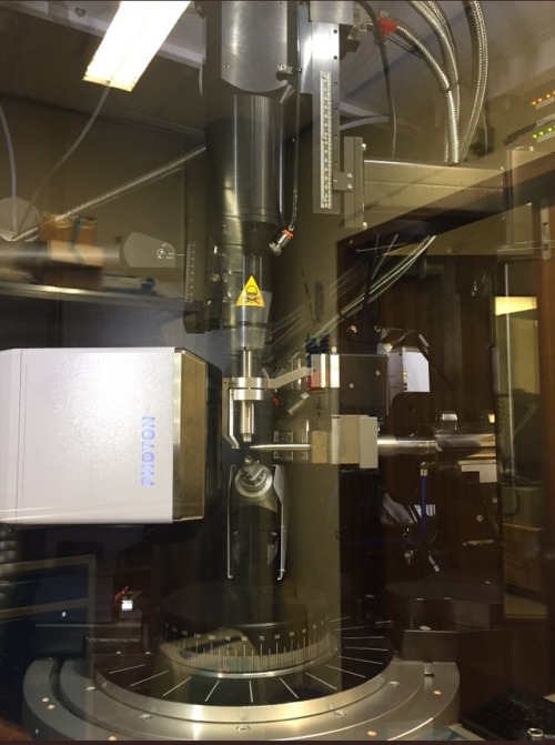
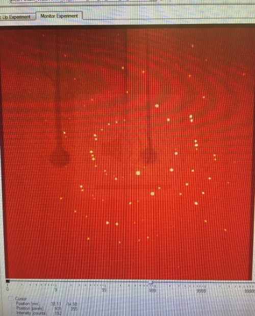
One of the in house crystallographers taught me how to use the X-ray diffractometer
XRD is awesome!
Post link
Befriend the crystallographers, they are wonderful people


