#forensic anthropology

I am setting up an internship to learn hands-on the technique of forensic facial reconstruction. The person teaching me works with the American Museum of Natural History (AMNH) here, in NYC.
The technique (anatomical method) is said to be more accurate than the tissue depth markers (alone), and computer. The anatomical or morphoscopic method for facial reconstruction involves rebuilding the face, muscle by muscle, tissue by tissue. This method is attributed to a Russian anthropologist, Mikhail M. Gerasimov (1907 - 1970), and it was the first technique developed in forensic sculpture. Gerasimov studied the face of more than 200 people and other human ancestors (Neanderthal and Java Man, 1927).

Image Source: Ullrich, Herbert & Stephan, Carl. (2016). Mikhail Mikhaylovich Gerasimov’s Authentic Approach to Plastic Facial Reconstruction. Anthropologie (Czech Republic). 54. 97-107.
The anatomical method is basically “fleshing out” the skull, meaning that piece by piece, glands, cartilage, and muscles are sculpted. The skull provides information “written” on the surface of the bone, whether it be muscle origin(s)/insertion points, as well as areas of smoothness where cartilage sits, or depressions where muscles lay. All of this combined with anatomy allows a face to be reconstructed from a skull, this then provides an identity for human remains.
Gerasimov, in the 1930s, realized that he was able to reconstruct faces of racial types (ancestries). Between 1937 - 1939, Gerasimov reconstructed the faces of a Papuan, Kazakh, and Khevsur Caucasian. Gerasimov also performed many forensic reconstructions for the NKVD. When Gerasimov reconstructed the faces of Yaroslav I the Wise (1938) and Andrei Bogolyubsky (1939) he received public attention. During WW2, Gerasimov worked at a hospital in Tashkent, and the various deceased victims worked as test subjects for statistical data on human skulls.

Image Source: Ullrich, Herbert & Stephan, Carl. (2016). Mikhail Mikhaylovich Gerasimov’s Authentic Approach to Plastic Facial Reconstruction. Anthropologie (Czech Republic). 54. 97-107.
Gerasimov honed his skills, and in 1950 received the USSR State Prize. Studying, and being interested in prehistoric animal bones as a child proved vital for Gerasimov’s craft. When the Soviet Ministry of Culture opened the tomb of Ivan the Terrible, Gerasimov reconstructed his face and received payment for his work. Gerasimov even traveled to Europe to find the skull of the poet, Schiller, within a mass grave. Gerasimov was not a man of politics and was always willing to help his friends.
Legacy: Gerasimov’s method of facial reconstruction spread all over the world. The anatomical method has been used to reconstruct the faces of pharaohs, and in the 1990s Russia used it to clarify the identities of the remains of the family of the last Tsar. You can see Gerasimov’s work at the State Historical Museum in Moscow, Anthropological Museum of Moscow State University, Museum of Georgia, and Museum of Uzbekistan.
Body size, scaling body size, and how shape changes.
Helps in a forensic context for the biological profile.
Population, and environmental stressors like mobility, and disease affect body size/shape. Can also be used to understand the sexual dimorphism of extinct hominins.
Ecogeographical rules related to thermoregulation:
Bergmann’s Rule (1847) If you have a species that is variable, and spread out over a geographical area you will see a larger variance of those species. Relating to body mass; bigger in colder climates, smaller in warmer climates.
Allen’s Rule (1877) concerned with appendage’s. Shorter in colder climates, and longer in warmer climates.
Together they are Bergmann’s and Allen’s rules.
A great modern example is observed in the following populations:
Inuit, Shorter limbs, and rounder stocky body:

Image Source: Ansgar Walk. Traditional clothing; left: seal, right: caribou (Iglulik). Wikimedia Commons.
Maasai, Longer limbs, slender taller body:
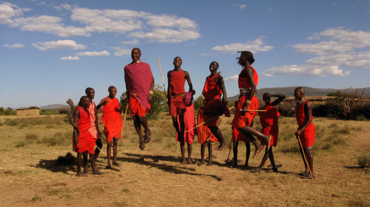
Image Source: Brutere. Maasai men performing traditional jumping dance (Adumu). Wikimedia Commons.
This is showing basic phenotypical adaptations to different climates, and how the environment can biologically change populations.
Note: Diet, nutrition, humidity, and variation (and many other variables) play a role in phenotype.
Forensic Friday: Sexual Dimorphism and Sexing

Image Source: Mike Peel. www.mikepeel.net. Yorkshire Museum - skeleton of a wealthy woman 1. Wikimedia Commons.
In 1901, during the construction of the York to Scarborough Railway Bridge, close to the River Ouse, workmen discovered a large stone coffin with a skeleton inside. The skeleton was found with many unusual, and expensive artifacts. This is a significant discovery from Roman York. Studying the skeleton revealed that it belonged to a woman.

Image Source: Mike Peel. www.mikepeel.net. Yorkshire Museum - skeleton of a wealthy woman 2. Wikimedia Commons.
Sexual Dimorphism in Humans:
On average, men are 8% larger than women.
Anatomical differences between men, and women can be easily seen in some soft tissue, however, this is limited in the skeleton. The human skeleton shows subtle morphological differences between the skull, dentition, pelvis, and long bones of men and women. On average, female skeletal elements are smaller, and less robust than males.
Some areas of skeletal differences amongst human males and females are:
- Note: variation, and these traits are basically nonexistent on pre-pubescent skeletal remains.
Skull: Mastoid processes, median nuchal line, supraorbital margin, supraorbital ridge, chin, and gonial angle of the mandible.
Dentition: Canines.
Pelvis: Greater sciatic notch, sub-pubic angle, sacrum, and pelvic cavity. Women have wider pelvises to allow for child birth.
LongBones: On average are longer, and have more pronounced muscle attachments in males.
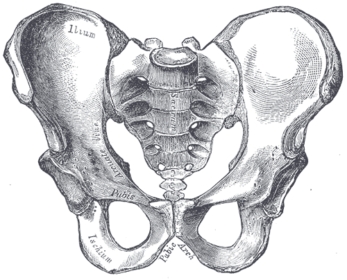
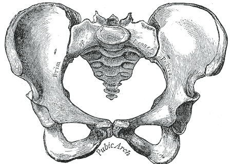
Image Source: Henry Vandyke Carter. Gray241. Wikimedia Commons.
Image Source: Henry Vandyke Carter. Gray242. Wikimedia Commons.
Left: Male pelvis. Right: Female pelvis.
How many carpals (wrist bones) are in one human wrist?
Can you name them all?
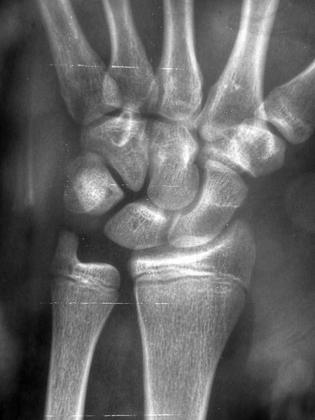
Image Source: Nevit Dilmen. Radiology 1300294. Wikimedia Commons.
Hint: “Some Lovers Try Positions That They Cannot Handle.”
Pop Quiz
Name the bone(s)?
What pathology do you notice at the shaft/diaphysis?
(Answers below)
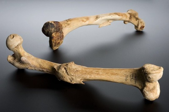
Image Source: Wellcome Images. Wellcome Trust. Human left femur, Tell Fara, Palestine, 100 BCE-200 CE Wellcome L0057387. Wikimedia Commons.
More information below:
“The femur bone is the bone joining the hip and knee joints. This femur shows an unreduced bone fracture. This means that the bones were not correctly realigned, using a splint or by surgical means, and therefore did not heal correctly. These femurs are from the left leg and the right leg of a human, although it is not clear if they are a pair. The bones were excavated at Tell Fara, Palestine, by the British School of Archaeology in Egypt, 1928-1929. The bones were purchased by Henry Wellcome from University College London the following year.”
It is past 3AM, and I am browsing through Amazon when…
Who is Phineas P. Gage (1823 - 1860)?
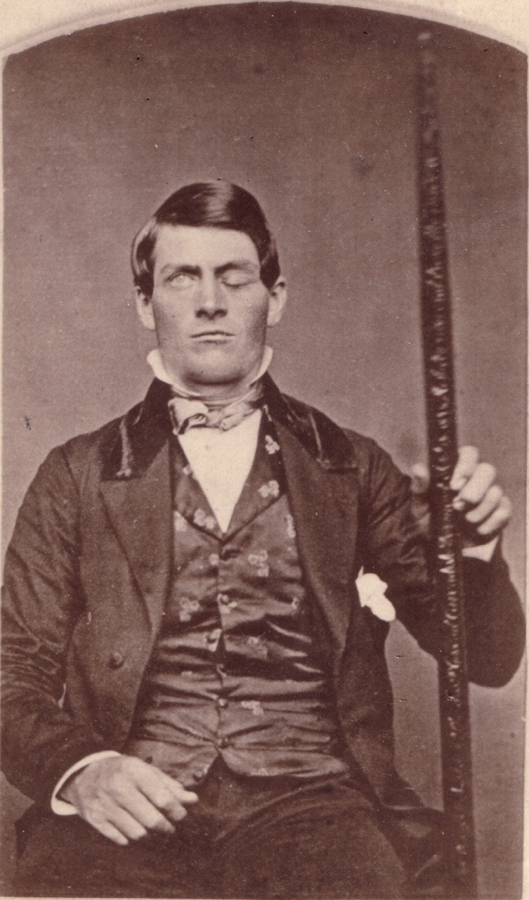
Image Source: Phineas Gage GageMillerPhoto2010-02-17 Unretouched Color Cropped. Public Domain. Wikimedia Commons.
Phineas P. Gage (1823–1860) was an American railroad construction foreman remembered for surviving an accident in which a large iron rod went completely through his skull. The iron rod destroyed a lot of Gage’s left side frontal lobe. After the injury, Gage lived for 12 years with effects on his personality, his friends described him as, “no longer Gage”.
The image below shows multiple views of the exhumed skull, and tamping iron, of Phineas Gage.

Image Source: J.B.S. Jackson, MD. JacksonJBS A descriptive catalogue of the Warren Anatomical Museum 1870 frontispiece 623x1024. A Descriptive Catalog of the Warren Anatomical Museum (1870). Wikimedia Commons.
Forensic Friday: Trepanation (or trepanning) is a surgical intervention in which a hole is drilled or scraped into the human cranium. The hole is made to expose the dura mater as a way to treat intracranial diseases, or to release pressure.

Image Source: Rama. Trepanated skull of a woman-P4140363-black. Wikimedia Commons.
Trepanation is one of the oldest surgical procedures with archaeological evidence.
Many prehistoric, and premodern patients had signs of bone healing, this means that many of these patients survived the surgery. Most of the crania with trepanation belong to adult males, but women and children also had the procedure done. In the New World, trepanation is commonly found among the Andean civilizations, and also in Mesoamerica.

Image Source: Wolfgang Sauber. Monte Albán - Trepanierter Schädel 2. Wikimedia Commons.
POP QUIZ:
Description of image: “Skull showing gunshot trauma Male profile, 1950s.”

Image Source: National Institutes of Health, Health & Human Services. Gunshot skull. Wikimedia Commons.
- Is this an exit, or an entrance wound? And why?
- Name the bone (or bones) affected by the gunshot trauma?
- Name two more bones observed in this image?
- What skeletal feature indicates this skull belongs to a male?
- Name one craniometric landmark observed in this image?
POP QUIZ!
How many bones do you see?
What joint is this?
Which side of the body are these bones from?

Image Source: Brian C. Goss. MedialHumerusRadiusUlnaArticulated. Wikimedia Commons.

Image Source: Sjbrown. Broken fixed arm. Wikimedia Commons.
Which two bones are fractured?

I am organizing for the coming academic semester, and I found a checklist/study guide from the last semester. Don’t forget to make studying fun! I’m a visual learner, and I use a lot of colors to make my notes more interesting to comprehend.
The Mayans developed great dental skills most likely for ritual, or religious purposes. It is believed that dental modifications were used to identify with a polity, ruler, region, or lineage. The Mayans placed carved stone inlays into prepared cavities on the front teeth. Minerals like jadeite, sepentine, pyrite, cinnabar, turquoise, hematite, and iron pyrites imbedded in these cavities can be observed in person at the Mexico City museum. To make the cavity on the enamel, the Mayans used a copper tube. The tube was spun between the hands, and powered quartz in water was incorporated as a means of making it abrasive. This action leads to the creation of a round hole on the tooth enamel. The stone inlays were grounded to fit exactly into the cavity. Younger children teeth have not been found with these modifications.

Image Source: Emmashavrick. Inlay dental modification. Wikimedia Commons.
Vocabulary: Latent Fingerprint
When deposits of oil and/or perspiration from a finger leaves a print on a surface it is known as a latent fingerprint. These fingerprints are not always visible to the naked eye, thus requiring a dusting technique with fine powders to make it visible. Once the power is on the oil/perspiration, it can be lifted with transparent tape for analyzing, protecting, and storing. Another technique for revealing latent fingerprints is to shine a laser at it, the laser will make the print(s) glow allowing for photographs to be taken.
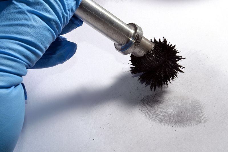
Image Source: Daekow. Fingerprint-magnabrush. Wikimedia Commons.
Aluminium is the most common powder used to develop latent fingerprints. A magna brush is a great tool for applying magnetic-sensitive powders over a hard surface. The aluminium adheres to the residue left behind by the oil/perspiration of a finger/hand, and reveals the print(s).

Image Source: National Institute of Standards and Technology (NIST). Latent Fingerprints (6774265822). Wikimedia Commons.
Fingerprints have been used for various purposes throughout human history. Thumb prints were used on clay seals in ancient China, and the ancient Babylonians used fingerprints on clay tablets for transactions.

Image Source: Mississippi Department of Archives and History. 6.13, 1963. Rifle that killed Medgar Evers. Located latent fingerprints on telescopic site. Medgar was shot off Delta Drive, Jackson, Miss. Wikimedia Commons.
Medgar Wiley Evers was an African American civil rights activist in Mississippi, and a World War Two veteran. On June 12, 1963, Medgar was assassinated by Byron De La Beckwith a member of the White Citizens’ Council, that was formed in Mississippi to resist the integration of schools, and civil rights activism. Medgar Wiley Evers was laid to rest with full military honors at Arlington National Cemetery. In the 1960s, the all-white juries failed to reach verdicts for the first two trials of Beckwith causing civil rights protests over the murder of Evers, and its trials. In 1994, new evidence in a new state trial led to Beckwith’s conviction
What is an osteosarcoma?
Who: Young woman, early 20s.
What: A malignant tumor of bone that has a rapid increase in osteoblasts (bone forming cells). The tumor measures at 24x23x19 centimeters, and was examined using x-rays, CT, and epi-illumination microscopy. The five pound tumor appears to not have affected the young woman’s food intake. The mandible is a rare location for such a tumor to develop.
When: 17th Century, unearthed in 1963.
Where: Buffalo, West Virginia, USA.
Osteosarcoma is the most common type of bone cancer, it mainly affects teens and young adults. For images about the case, take a look at the article below.
News Source: Basketball-Sized Jaw Tumor Found On Skeleton Of 17th Century Woman In West Virginia
Basketball-Sized Jaw Tumor Found On Skeleton Of 17th Century Woman In West Virginia
Life Update:
Hello everyone!
After a fun, and challenging first semester of grad school (and a short break from blogging) I have returned. On a great note, I am proud to say that I earned an “A” in the Human Osteology and Odontology course! This puts me one step closer to interning at the office of chief medical examiners in NYC.

My instructor is phenomal, and I learned to look a bone in such an interesting way. For those that might be interested, the course involved looking at bone fragments (not whole bone) and determining whether the bone is human or nonhuman, the element (what bone it is), where on the bone is the fragment from, side of the body the bone is from, and lastly whether or not the fragment came from an adult or a subadult human. We also looked at some important soft-tissue, as well as development, and pathologies. Over all, the course was a great experience filled with many hours in the osteology lab. Super grateful to have had such an opportunity.
Classes start again at the end of this month, and I will be taking three courses again. One is about the history of biological anthropology, another is an internship at the molecular anthropology lab (Super excited for this!), and Interpreting The Human Skeleton, the course taken after osteo.

Image Source: Frank E. Beddard. The Cambridge Natural History, Volume X, Mammalia. Wikimedia Commons.
Here is a drawing of a human atlas, also known as the first cervical vertebrae C1. The image above depicts a young/not fully fused atlas, this is shown by the development of the anterior arch of the vertebral foramen (ia) not yet fusing to the anterior portion near the articular surface of the occipital condyles (as). Other notable landmarks include; the transverse process (t)— the transfers foramen directly medial to it, and the groove where the first spinal nerve and vertebral artery go through (g). The atlas has no vertebral body, no articular discs on its superior or inferior, and lacks a spinous process. On the posterior and medial surface of ia is the articulation for the dens of the C2.
Below is a fully fused C1/Atlas.

Image Source: Gliu. Atlante, prima vertebra cervicale, vista superiore. Wikimedia Commons.
POP QUIZ: Why do you think the C1 was nicknamed “Atlas”?

Some drawings of the C2 and C5.
Pop Quiz: How many Cervical Vertebrae do humans usuallyhave?

Image Credit: Pasini et al., World Neurosurgery, 2018
A rare phenomenon of post mortem fetal extrusion (coffin birth).
The remains are of a 38 week old fetus found in a grave dated between the 7th and 8th century in the town of Imola, Bologna, Italy. The woman had been purposely buried, and also shows markings on her skull that indicate she had undergone mideval brain surgery at least one week before her death.
“Coffin Birth” also known as post-mortem fetal extrusion is a rare phenomenon. In a case of coffin birth, the fetus is expelled from a pregnant woman’s body after she has died (the fetus are also deceased or die). When humans decompose, gases are produced and internal pressure builds. Pressure is placed on the internal organs and sometimes forces a fetus through the vaginal canal.

Image Credit: Pasini et al., World Neurosurgery, 2018
Fact: The earliest recorded case of coffin birth was in 1551. The Spanish Inquisition ordered a woman to be hanged by the neck, four hours later two dead babies were seen falling from her body.

Image Source: Didier Descouens. human skull. Wikimedia Commons.
A human cranium gets cut open (like the one pictured above) to remove the brain, either for science or legal purposes.