#popquiz

Pop Quiz
Name the bone(s)?
What pathology do you notice at the shaft/diaphysis?
(Answers below)
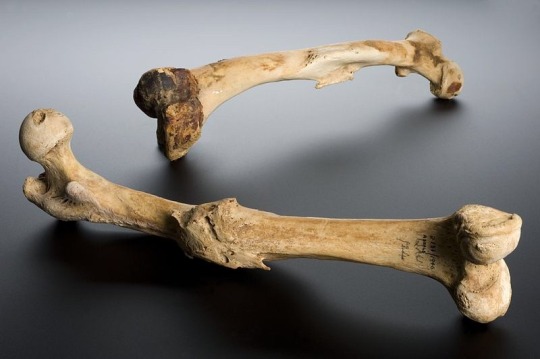
Image Source: Wellcome Images. Wellcome Trust. Human left femur, Tell Fara, Palestine, 100 BCE-200 CE Wellcome L0057387. Wikimedia Commons.
More information below:
“The femur bone is the bone joining the hip and knee joints. This femur shows an unreduced bone fracture. This means that the bones were not correctly realigned, using a splint or by surgical means, and therefore did not heal correctly. These femurs are from the left leg and the right leg of a human, although it is not clear if they are a pair. The bones were excavated at Tell Fara, Palestine, by the British School of Archaeology in Egypt, 1928-1929. The bones were purchased by Henry Wellcome from University College London the following year.”
POP QUIZ:
Description of image: “Skull showing gunshot trauma Male profile, 1950s.”

Image Source: National Institutes of Health, Health & Human Services. Gunshot skull. Wikimedia Commons.
- Is this an exit, or an entrance wound? And why?
- Name the bone (or bones) affected by the gunshot trauma?
- Name two more bones observed in this image?
- What skeletal feature indicates this skull belongs to a male?
- Name one craniometric landmark observed in this image?
POP QUIZ!
How many bones do you see?
What joint is this?
Which side of the body are these bones from?

Image Source: Brian C. Goss. MedialHumerusRadiusUlnaArticulated. Wikimedia Commons.

Image Source: Frank E. Beddard. The Cambridge Natural History, Volume X, Mammalia. Wikimedia Commons.
Here is a drawing of a human atlas, also known as the first cervical vertebrae C1. The image above depicts a young/not fully fused atlas, this is shown by the development of the anterior arch of the vertebral foramen (ia) not yet fusing to the anterior portion near the articular surface of the occipital condyles (as). Other notable landmarks include; the transverse process (t)— the transfers foramen directly medial to it, and the groove where the first spinal nerve and vertebral artery go through (g). The atlas has no vertebral body, no articular discs on its superior or inferior, and lacks a spinous process. On the posterior and medial surface of ia is the articulation for the dens of the C2.
Below is a fully fused C1/Atlas.

Image Source: Gliu. Atlante, prima vertebra cervicale, vista superiore. Wikimedia Commons.
POP QUIZ: Why do you think the C1 was nicknamed “Atlas”?

Some drawings of the C2 and C5.
Pop Quiz: How many Cervical Vertebrae do humans usuallyhave?
Riddle:
Which came first, the hamulus or the hamate?

Image Source: Brian C. Goss. Photograph of right anterior human distal radius and ulna. Keywords: Human bones, bones, wrist, radius, ulna. Wikimedia Commons.
Pop Quiz!

Image Source: BodyParts3D is made by DBCLS. Tarsal Bones. Wikimedia Commons.
Name the bones based on their shade/color:
- (Within parenthesis in standard anatomical position you will find the directional terms of the feet shown above)
Yellow (most posterior):
Lavender (most superior):
Violet (most lateral):
Red (most medial):
BONUS: How many cuneiform bones are in one human foot?
ClickHERE for the answers.