#osteology

Forensic Friday: Sexual Dimorphism and Sexing

Image Source: Mike Peel. www.mikepeel.net. Yorkshire Museum - skeleton of a wealthy woman 1. Wikimedia Commons.
In 1901, during the construction of the York to Scarborough Railway Bridge, close to the River Ouse, workmen discovered a large stone coffin with a skeleton inside. The skeleton was found with many unusual, and expensive artifacts. This is a significant discovery from Roman York. Studying the skeleton revealed that it belonged to a woman.

Image Source: Mike Peel. www.mikepeel.net. Yorkshire Museum - skeleton of a wealthy woman 2. Wikimedia Commons.
Sexual Dimorphism in Humans:
On average, men are 8% larger than women.
Anatomical differences between men, and women can be easily seen in some soft tissue, however, this is limited in the skeleton. The human skeleton shows subtle morphological differences between the skull, dentition, pelvis, and long bones of men and women. On average, female skeletal elements are smaller, and less robust than males.
Some areas of skeletal differences amongst human males and females are:
- Note: variation, and these traits are basically nonexistent on pre-pubescent skeletal remains.
Skull: Mastoid processes, median nuchal line, supraorbital margin, supraorbital ridge, chin, and gonial angle of the mandible.
Dentition: Canines.
Pelvis: Greater sciatic notch, sub-pubic angle, sacrum, and pelvic cavity. Women have wider pelvises to allow for child birth.
LongBones: On average are longer, and have more pronounced muscle attachments in males.
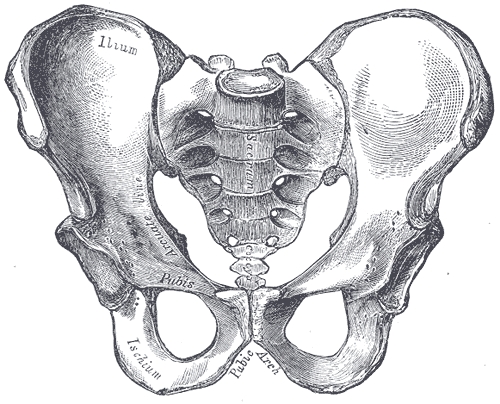
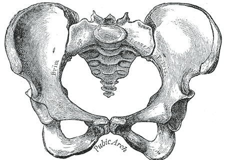
Image Source: Henry Vandyke Carter. Gray241. Wikimedia Commons.
Image Source: Henry Vandyke Carter. Gray242. Wikimedia Commons.
Left: Male pelvis. Right: Female pelvis.
How many carpals (wrist bones) are in one human wrist?
Can you name them all?
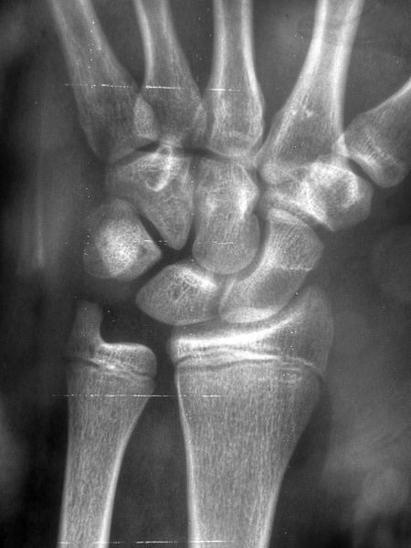
Image Source: Nevit Dilmen. Radiology 1300294. Wikimedia Commons.
Hint: “Some Lovers Try Positions That They Cannot Handle.”
Pop Quiz
Name the bone(s)?
What pathology do you notice at the shaft/diaphysis?
(Answers below)
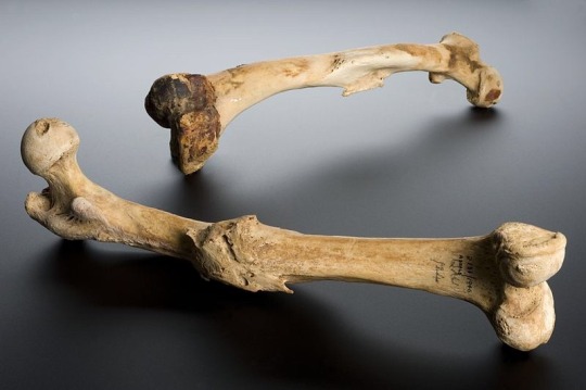
Image Source: Wellcome Images. Wellcome Trust. Human left femur, Tell Fara, Palestine, 100 BCE-200 CE Wellcome L0057387. Wikimedia Commons.
More information below:
“The femur bone is the bone joining the hip and knee joints. This femur shows an unreduced bone fracture. This means that the bones were not correctly realigned, using a splint or by surgical means, and therefore did not heal correctly. These femurs are from the left leg and the right leg of a human, although it is not clear if they are a pair. The bones were excavated at Tell Fara, Palestine, by the British School of Archaeology in Egypt, 1928-1929. The bones were purchased by Henry Wellcome from University College London the following year.”
It is past 3AM, and I am browsing through Amazon when…
POP QUIZ!
How many bones do you see?
What joint is this?
Which side of the body are these bones from?

Image Source: Brian C. Goss. MedialHumerusRadiusUlnaArticulated. Wikimedia Commons.

I am organizing for the coming academic semester, and I found a checklist/study guide from the last semester. Don’t forget to make studying fun! I’m a visual learner, and I use a lot of colors to make my notes more interesting to comprehend.

Some drawings of the C2 and C5.
Pop Quiz: How many Cervical Vertebrae do humans usuallyhave?

Image Credit: Pasini et al., World Neurosurgery, 2018
A rare phenomenon of post mortem fetal extrusion (coffin birth).
The remains are of a 38 week old fetus found in a grave dated between the 7th and 8th century in the town of Imola, Bologna, Italy. The woman had been purposely buried, and also shows markings on her skull that indicate she had undergone mideval brain surgery at least one week before her death.
“Coffin Birth” also known as post-mortem fetal extrusion is a rare phenomenon. In a case of coffin birth, the fetus is expelled from a pregnant woman’s body after she has died (the fetus are also deceased or die). When humans decompose, gases are produced and internal pressure builds. Pressure is placed on the internal organs and sometimes forces a fetus through the vaginal canal.

Image Credit: Pasini et al., World Neurosurgery, 2018
Fact: The earliest recorded case of coffin birth was in 1551. The Spanish Inquisition ordered a woman to be hanged by the neck, four hours later two dead babies were seen falling from her body.

Image Source: Didier Descouens. human skull. Wikimedia Commons.
A human cranium gets cut open (like the one pictured above) to remove the brain, either for science or legal purposes.
Name one bone of the skull that ossifies endochodrally? (Cartilaginous Precursor)

One of the journal sketches I completed today.

Pop Quiz!
What is the name the type of vertebrate shown above?
- A. Cervical
- B. Thoracic
- C. Lumbar
- D. Sacrum
Bonus: What number within said specific vertebral category is the vertebra shown above?
For my osteology class I must hand in a sketchbook.
I am not the best at drawing, I took ceramics during undergrad because it was the cheapest art requirement course. If you are not the best at drawing I recommended you practice! Drawing is a great way to learn Osteology.
Riddle:
Which came first, the hamulus or the hamate?

Image Source: Brian C. Goss. Photograph of right anterior human distal radius and ulna. Keywords: Human bones, bones, wrist, radius, ulna. Wikimedia Commons.
Pop Quiz!

Image Source: BodyParts3D is made by DBCLS. Tarsal Bones. Wikimedia Commons.
Name the bones based on their shade/color:
- (Within parenthesis in standard anatomical position you will find the directional terms of the feet shown above)
Yellow (most posterior):
Lavender (most superior):
Violet (most lateral):
Red (most medial):
BONUS: How many cuneiform bones are in one human foot?
ClickHERE for the answers.

Image Source: Arielinson. CT scan converted into an animated 3d model using Phodtoshop. Human skull. Wikimedia Commons.

Tumbas de Tiro
En las Tumbas de Tiro se depositaban difuntos de alto rango junto con ofrendas que facilitarían su tránsito al mundo de los muertos. Conoce como se llevaba a cabo esta práctica en la sala Culturas de Occidente del Museo Nacional de Antropología.
The Western Mexico shaft tomb tradition or “shaft tomb culture” where the high ranking departed were buried and placed with offerings (ofrendas) to help them transition to the world of the dead (Mundo de los muertos). This tradition is thought to have started around 300 BCE. The bodie(s) sometimes had these items next to them: hollow ceramic figures, obsidian and shell jewelry, semi-precious stones, and pottery, which often had food inside.

Image Source: Maunus. Shaft tomb exhibited at the Museo Nacional de Antropología e Historia, México. Wikimedia Commons.
Post link


Started this one out in a workshop with the mighty Kathryn Engberg, I added some extra sessions on my own and dropped a skeleton to study the pose. The triceps, teres major, teres minor and gluteus minimus are there just because I like them.
Post link


 Upper Cave China 101+103, Mladec 1 Czech Republi")

Almost 6,000 years ago, the man was placed behind the woman with his arms around her body, and their legs were intertwined. They were buried.
Why they were interred in this manner is not yet determined, but the international team that discovered them in Greece is still searching for answers, according to team member Michael Galaty, a Mississippi State University archaeologist.
“There’ve only been a couple of prehistoric examples of this behavior around the world, but even when couples are buried together, they’re beside each other and not typically touching,” he said. “This couple was actually spooning. We assume they were partners of some kind, and because of DNA analysis, we do know they are male and female.”
Not only does Galaty head MSU’s anthropology and Middle Eastern cultures department, but he also serves as interim director of the university’s Cobb Institute of Archeology.
Another question for the researchers to examine is how the couple died, which happened around 3800 B.C., Galaty said. While archaeologists are unsure whether the man or woman died first, they are sure the couple’s times of death are close together.
“This is unique in Greece, and we’re analyzing the skeletons and bones to find out more about what was going on, how they died and why they may have been placed there,” he said.
Post link