#microtubules
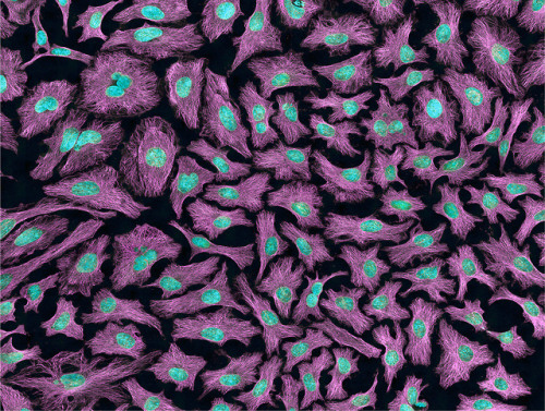
Multiphoton fluorescence image of HeLa cells with cytoskeletal microtubules (magenta) and DNA (cyan). Nikon RTS2000MP custom laser scanning microscope.
Source: National Center for Microscopy and Imaging Research (NCMIR) (source)
Credit Line: National Center for Microscopy and Imaging Research
Post link
Image of the Week – June 8, 2020
CIL:12375 -http://cellimagelibrary.org/images/12375
Description: Movie showing the dynamics of kinetchore microtubules during meiosis II in primary spermatocytes of the crane-fly Nephrotoma suturalis that were experimentally flattened. Time-lapse polarization microscopy using a Nikon Microphot SA, equipped for liquid crystal polarized light microscopy (LC-PolScope, CRi, Woburn Massachusetts) 60x/1.4 PlanApo oil immersion objective, 1.4 NA oil imm. condenser, with 2.0x zoom lens. Images captured every 15 sec using a QImaging Retigo EXi CCD camera. Raw images were processed using 5-frame algorithm (Shribak and Oldenbourg, 2003). The time series used for the movie is included in this grouped set.
Authors: James R. LaFountain and Rudolf Oldenbourg
Licensing: Public Domain: This image is in the public domain and thus free of any copyright restrictions. However, as is the norm in scientific publishing and as a matter of courtesy, any user should credit the content provider for any public or private use of this image whenever possible.

