#cell biology

In really surreal science news of the day, researchers have now found a way to watch how hundreds of individual cells work together to maintain and regenerate skin tissue, thanks to a genetically engineered line of technicolor zebrafish.
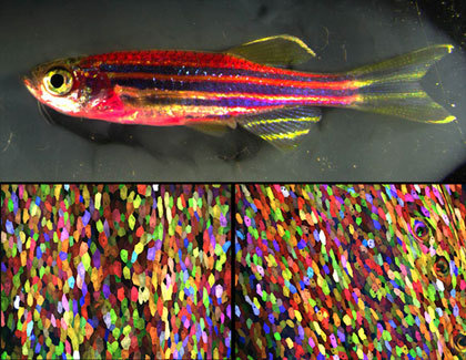
(To the naked eye, this genetically engineered zebrafish has a magenta tone (top). But under a microscope, every cell has a distinct color, thanks to a new labeling technique called Skinbow (bottom). Photo credit: Chen-Hui Chen, Duke University)
Ken Poss, a professor of cell biology at Duke University, and his lab have spent the last few years creating “Skinbow,” a mutant zebrafish with fluorescent markers embedded in the DNA code of its skin cells. Their study was published on March 21st, 2016 in Developmental Cell, and is available for free download: Multicolor Cell Barcoding Technology for Long-Term Surveillance of Epithelial Regeneration in Zebrafish.
Each of Skinbow’s color markers can be expressed in three different ways, resulting in red, green, or blue fluorescence. With three color options and roughly a hundred copies of the gene per cell, the total number of colors any cell can take on is probably in the thousands.
The research team has now been able to observe tissue regeneration by scraping a few cells off the fish’s fin, and visualize the healing process on a cellular level. Any color patterns that develop during tissue regeneration are inferred to arise from a single cell.
“It is like you have given each cell an individual barcode,”said Chen-Hui Chen, a postdoctoral fellow in Poss’s lab and lead author on the study. “You can precisely see how individual cells collectively behave during regeneration.”
Listen to Ken Poss discuss the experiment:
“One thing we weren’t expecting is that within a few hours of injury, cells that are spared acquire some mobility on the surface,”Posssaid in an interview with Gizmodo. “They also expand in size, some doubling. Then there’s a quick wave of replacement—you can see new cells emerging from the layer underneath.”
What is even more fascinating is that cells behaved differently in function of the injury. For example, following fin amputation, pre-existing cells were initially recruited from below the amputation plane to cover the wound. Then, new cells were generated at a rapid pace, and in the end some cells at the tip of the regrown tissue temporarily grew in size to provide surface coverage.

(Here, the multicolor cells are evident on the exposed portion of a dissected scale. Photo credit: Chen-Hui Chen, Duke University)
Poss is hopeful that the Skinbow technique can be extended to other species in the future, even humans. It obviously would involve getting the right set of genetic tools, and developing a way to image tissue continually over time, but Poss is hopeful that doctors may be able to use this technology to visualize how human tissue responds to new cancer drugs, or treatments meant to accelerate healing.
 and #nuclei using DAPI (green). T")
#mitochondria stained using #immunofluorescence for TOM20 (orange) and #nuclei using DAPI (green). To show the morphology of MEF cells, they used bright field #microscopy (detection with TPMT) . For confocal #imaging they utilized a @zeiss_micro LSM 710 at the LMF @DZNE_en.
Image courtesy of Christian Lamberg, PhD (@Christi23003438 on twitter) (see original post)
Post link

Colony of stem cells derived from an individual with bipolar disorder
Source : medicine.umich.edu
Post link

 and DNA (cyan)")
Multiphoton fluorescence image of HeLa cells with cytoskeletal microtubules (magenta) and DNA (cyan). Nikon RTS2000MP custom laser scanning microscope.
Source: National Center for Microscopy and Imaging Research (NCMIR) (source)
Credit Line: National Center for Microscopy and Imaging Research
Post link

Fig. 4. Overexpression of LGP85 does not cause enlargement of lysosome, but results in formation and accumulation of the late endosome-lysosome hybrid organelle. To label lysosomes, COS cells were incubated with Texas-Red dextran for 4 hours and chased for 20 hours. After that, cells were transiently transfected with LGP85 and fixed at 12 (A-C), 24 (D-F), and 36 (G-I) hours after transfection, followed by staining for LGP85 (A,D,G, green). Cells were visualized by confocal microscopy. The right columns show the merged images of LGP85 (green) and dextran (red). Bars, 20 μm.
A role for the lysosomal membrane protein LGP85 in the biogenesis and maintenance of endosomal and lysosomal morphology
Toshio Kuronita, Eeva-Liisa Eskelinen, Hideaki Fujita, Paul Saftig, Masaru Himeno, Yoshitaka Tanaka
Journal of Cell Science 2002 115: 4117-4131; doi: 10.1242/jcs.00075
Post link


#Lipilight#MemBright and super resolution #LiveSR on living muscle cells! by Bruno Cadot
Visit website : https://cadotbruno.com
Visit Bruno’s Twitter : https://twitter.com/cadotbru
Post link

Stem cells in the skin - stem cells labelled in red are found in special microenvironments where they are surrounded by other types of cell.
Post link

A rendered image of a primary neuronal stem cell culture in which cells were labeled with different fluorescently labeled proteins that differentiate between stem cells (orange/yellow) and their neuronal ‘offspring’ (blue/ green/ purple).
Post link
, vinc")
Huntington’s stem cell derived oligodendrocyte precursors stained for phalloidin (green), vinculin (red) and DNA (blue).
Post link

 and Nissl bodies (green).")
Rat dorsal root ganglia cells stained for Alpha Acetyl tubulin (red) and Nissl bodies (green).
Post link
, GAD65 (green) and DNA (blue).")
Trisomy 21 derived neural cells stained for neurofilament heavy (red), GAD65 (green) and DNA (blue).
Post link

Mouse diaphragm consisting of neurons, muscle cells and neuromuscular junctions under the confocal microscope: Green structures show neurons (Alexa 488), red areas are neuromuscular junctions (rhodamin), and blues areas are muscle fiber (myosin, DODT contrast).
Post link

Human epithelial cell in mitosis, fluorescently labeled for alpha tubulin, gamma tubulin and DNA.
Post link


 and now something for this occasion:Photomicrogr")
Nearly 2K followers on tumblr! wtf is happening? ;) and now something for this occasion:
Photomicrograph of a midsaggital section showing the different components of the rat cerebellum, including Purkinje neurons in green, glia (non-neuronal cells) in red, and cell nuclei in blue. This image is part of an ongoing effort to develop methods to enable the creation large-scale digital atlases of the brain.
Post link

Image shows adult human fibroblast cells with intracellular proteins involved in adhesion of these cells to an extracellular matrix. Magenta represents actin stress fibers in a cell and green staining represents a focal adhesion protein vinculin, which together contribute to how strongly these cells adhere to a matrix surface. Blue is the nucleus of a cell.
Post link

Leishmania major promastiotes during infection of primary fibroblast culture. Cells are stained with anti-tubulin (green) and anti-actin (red).
Post link
 and blood vessels (green) | ZEISS Microscopy A mouse’s fat cells (red) are sho")
A mouse’s fat cells (red) are shown surrounded by a network of blood vessels (green). Fat cells store and release energy, protect organs and nerve tissues, insulate us from the cold and help us absorb important vitamins. Image courtesy of Daniela Malide, National Heart, Lung, and Blood Institute, National Institutes of Health.
Post link