#microscopy
Battery: In-situ Microscopy
Nano-microscope gives first direct observation of the magnetic properties of 2D materials
Australian researchers and their colleagues from Russia and China have shown that it is possible to study the magnetic properties of ultrathin materials directly, via a new microscopy technique that opens the door to the discovery of more two-dimensional (2D) magnetic materials, with all sorts of potential applications.
Published in the journal Advanced Materials, the findings are significant because current techniques used to characterise normal (three-dimensional) magnets don’t work on 2D materials such as graphene due to their extremely small size – a few atom thick.
“So far there has been no way to tell exactly how strongly magnetic a 2D material was,” said Dr Jean-Philippe Tetienne from the University of Melbourne School of Physics and Centre for Quantum Computation and Communication Technology.
“That is, if you were to place the 2D material on your fridge’s door like a regular fridge magnet, how strongly it gets stuck onto it. This is the most important property of a magnet.”
To address the problem, the team, led by Professor Lloyd Hollenberg, employed a widefield nitrogen-vacancy microscope, a tool they recently developed that has the necessary sensitivity and spatial resolution to measure the strength of 2D material.
Spooky science
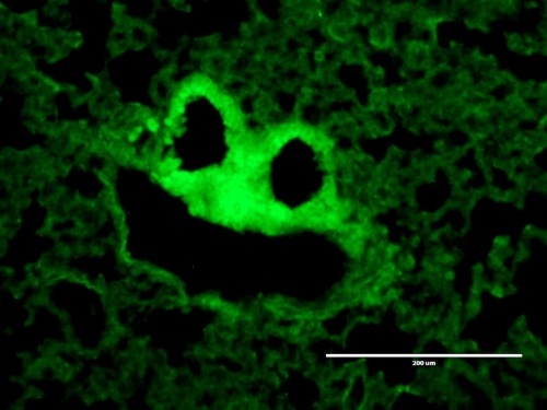
This ghoulish image shows lung tissue structure.
Laura Sibley who took this image is part of a team from Royal Holloway, who are developing novel vaccines using bacteria (similar to ones that are used in probiotic drinks).
The vaccines they are creating are cheaper than normal vaccines, easier to produce and have no chemicals in them, making them suitable for diseases affecting developing countries. There are many diseases that need vaccine development, including tuberculosis, which kills around two million people per year, and is one of the diseases that they are focused on.
Laura stumbled across the ghostly vision in a study looking at TB vaccine distributed in lung tissue, work which in the future could help to protect people against TB infection.
Post link
24 hours in the life of a zebrafish embryo
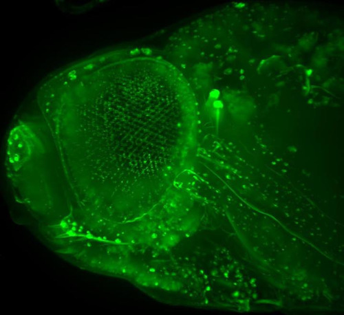
In this video you can watch 24 hours (the second day) in the development of a tiny zebrafish embryo.
Some cells have been labelled with a fluorescent protein naturally found in jellyfish. This fluorescent label marks several developing organ systems including the eye, hatching gland and kidney.
You can also see two groups of cells migrating along the trunk of the embryo, depositing smaller clumps of sensory cells as they go. These cells are the precursors of the lateral line system, which gives fish a sense of ‘touch at a distance’, enabling them to shoal, avoid obstacles and find prey.
Look more closely to see individual cells moving around over the skin.
Light sheet microscopy illuminates the specimen with a thin sheet of light and allows scientists like Dr Tanya Whitfield and her team at the University of Sheffield to observe cells and embryos at high speeds or for long times. This research is helping them to gain a better understanding of the amazing process of embryogenesis, including the development of sensory systems such as the lateral line and inner ear.
Video: Sarah Baxendale and Nick van Hateren.
See more beautiful images of zebrafish embryos here
A two-day-old zebrafish embryo brain
These striking images show a zebrafish embryo brain at just two-days-old as seen with a Light Sheet Fluorescent Microscope.
Light sheet microscopy illuminates the specimen with a thin sheet of light and can be used to see relatively large structures in fine detail. You can see all the nerve cells and connections in the zebrafish brain stained with an antibody in red.
Being able to see embryos in this detail allows research teams at the University of Sheffield to follow the amazing process of embryogenesis, from a single cell (the fertilised egg) to a functioning animal with hundreds of different cell types.
Images: Sarah Baxendale, Stone Elworthy and Nick van Hateren.
Post link
Flies offer insights into motor neuron disease
ALS is one of the most common forms of motor neuron disease, killing around 1,200 people a year in the UK.
Researchers at the Babraham Institute and the University of Massachusetts have developed a new way to study the process of nerve damage, which occurs in people with ALS and other forms of motor neuron disease.
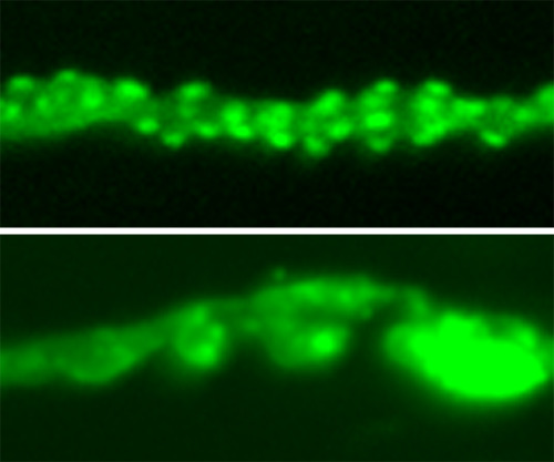
By studying nerves in legs from the common fruit fly (top image), researchers identified genes involved in the neurodegeneration process.
They hope that this model will both increase understanding of neurodegenerative diseases and help to inform future therapies.
Image credits: Top: Microscopic image of a fly leg, Babraham Institute
Bottom left: (top) fully-functioning neuromuscular junction from a wild type fly and neuromuscular junction showing degeneration (bottom), Babraham Institute
Bottom right: Drosophila pupae, Helio Alexandre Duarte Roque forZEISS Microscopy
Post link
Ciliated epithelial cell in a BAL sample.
Water flea
Discarded AFM tips.
Atomic Force Microscopy, or AFM, is a technique by which a small mechanical probe is scanned across a sample to create a height map. This technique has very high resolution, less than a nanometer, depending on what kind of tip is being used, and can be done in ambient conditions (no need for vacuum). AFM is useful for getting roughness data and measuring film thickness, and can be combined with other microscopy techniques to get a complete picture of your device.
AFM probes often get damaged or dirty, resulting in “tip graveyards” like the one shown here.
Post link
The past few weeks I have been working hard to produce a new collection of unique fine jewellery and objects.
Each piece starts in wax, which I carefully shape before it is cast into precious metal. I then meticulously solder and hand work each metal form before setting carefully selected diamonds, sapphires and other valuable gemstones. For this collection I have hand set over 100 gems
Visit @po8gallery this month to see the jewellery and objects from “beneath the surface” along with a series of large neuroscience prints I created while @qldbraininstitute
.
.
#goldpendant #wip #neuroscience #microscopy #beautifulscience #sciart #sciencejewelry #contemporaryjewellery #australiandesigner #organicjewelry #madebyhand #australiansapphire
Post link
Normally invisible, these dynamic branches and tubulations are a just a few of the many internal compartments of a single cell. They’ve been made to glow by using green fluorescent protein found in jellyfish
.
When I was studying neuroscience all I wanted to do was get involved in research on consciousness, but I took a side step and started an imaging project in a cancer biology lab with Prof. Jenny Stow @uniofqld This is when I first started using microscopes to image fluorescent proteins in cells.
Its one thing to know that without your awareness every single cell in your body is almost vibrating with activity and something entirely different to actually see it with your own eyes. Looking down into an ocean of darkness and seeing these glowing worlds alive within cells was a profound experience that completely captured my imagination. A few years later I started work establishing an imaging facility @qldbraininstitute so these techniques could be used to study the brain. It’s been such an incredible journey to see this technique move from allowing us to see inside a single cell, like in this movie I captured over a decade ago, to watching every neuron in a brain flashing with activity.
.
This dynamic intracellular activity and the even smaller structures that surround these tubules and spheres helped inspire the jewellery and objects I’m exhibiting with @po8gallery this month. Opening night Thursday 6th. Come and see!!
.
.
#neuroscience #microscopy #beautifulscience #sciart #microscopic #fluorescence #contemporaryjewellery #australiandesigner #organicjewelry #madebyhand
Setting blue green Australian sapphires, one of the final steps in creating one of the larger rings for ‘beneath the surface’ - opening @po8gallery this Thursday
.
.
.
#contemporaryjewellery #australiandesigner #organicjewelry #madebyhand #australiansapphire #neuroscience #microscopy #beautifulscience #scienceisart #sciart #wip #beneaththesurface
A number of the pieces I’ve created for the exhibition opening @po8gallery next week combine both lost wax and fabrication techiques. This ring holds a beautiful 1ct Australian parti sapphire in 18ct white gold. Illuminated from all sides and visible from above it sits protected by an organic veil of 18ct yellow gold.
.
Beneath the Surface explores our growing capability to make the invisible visible, which is revealing previously unknown worlds of hidden beauty within us.
.
.
#neuroscience #microscopy #beautifulscience #sciart #sciencejewelry #contemporaryjewellery #australiandesigner #organicjewelry #madebyhand #australiansapphire
Post link
Sneak preview for my upcoming solo show with @po8gallery / A selection of 11 Australian blue and green sapphires with a brilliant white diamond are set into the inner surface of this 18ct yellow gold ring. Ready for the final centre stone
.
Beneath the Surface opens April 6 and will include a new collection of fine jewellery, silver sculpture and digital images from my work @qldbraininstitute
.
#neuroscience #microscopy #beautifulscience #brain
#scienceisart #sciart #sciencejewelry #contemporaryjewellery #australiandesigner #organicjewelry #madebyhand #wip #beneaththesurface

Bender-scope
The microscopes of the future are all surly and a little bit hungover.
Current mood: “Bite my shiny metal a$$“
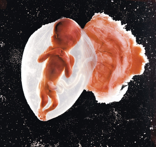
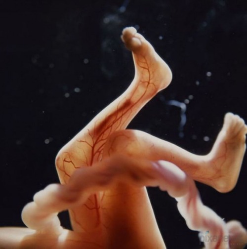
RIP Lennart Nilsson (1922-2017), the man who brought us some of our earliest and most vivid glimpses into human life in the womb.
Post link
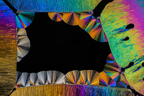
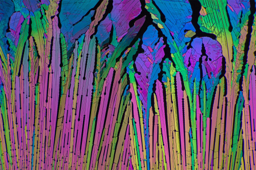
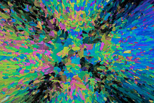
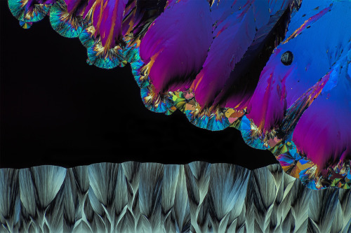
Painkillers are pretty. Or at least acetaminophen is when crushed, melted onto a slide and photographed by Peter Juzak
Post link
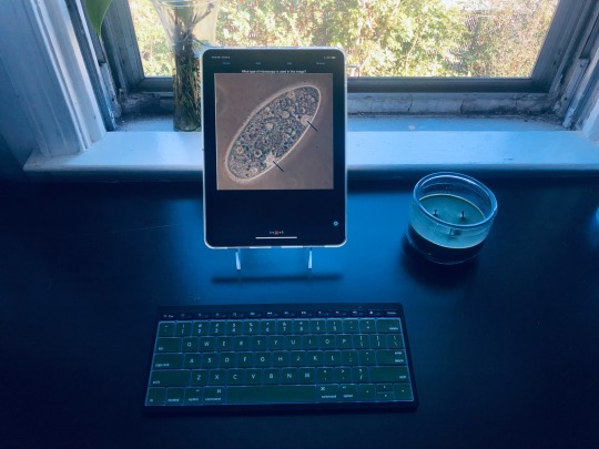
some anki flashcards for all your microscopy identification needs :) this anki deck is actually linked on my blog if you’re studying for a microbiology/intro bio course and need to learn how images appear under different microscopes!

