#anatomy


Human skeleton, 1732
These illustrations – created in 1732 for an article published in 1741 by an ophthalmologist in Kyōto named Toshuku Negoro – show the skeletal remains of two criminals that had been burned at the stake.

Human skeleton, 1732
This document is thought to have inspired physician Tōyō Yamawaki to conduct Japan’s first recorded human dissection.


Japan’s first recorded human dissection, 1754
These illustrations are from a 1754 edition of a book entitled Zōzu, which documented the first human dissection in Japan, performed by Tōyō Yamawaki in 1750. Although human dissection had previously been prohibited in Japan, authorities granted Yamawaki permission to cut up the body of an executed criminal in the name of science. The actual carving was done by a hired assistant, as it was still considered taboo for certain classes of people to handle human remains.


Edo-Period, Japan.
Breast cancer treatment, 1809.
These illustrations are from an 1809 book documenting various surgeries performed by Seishū Hanaoka for the treatment of breast cancer. The illustrations here depict the treatment for a 60-year-old female patient.
Post link
Here is a selection of old anatomical illustrations that provide a unique perspective on the evolution of medical knowledge in Japan during the Edo period (1603-1868).

Trepanning instruments, circa 1790
These illustrations are from a book on European medicine introduced to Japan via the Dutch trading post at Nagasaki. Pictured here are various trepanning tools used to bore holes in the skull as a form of medical treatment.

Trepanning instruments, circa 1790
The book was written by Kōgyū Yoshio, a top official interpreter of Dutch who became a noted medical practitioner and made significant contributions to the development of Western medicine in Japan.

Trepanning instruments, 1769
These illustrations of trepanning instruments appeared in an earlier book on the subject
Here is a selection of old anatomical illustrations that provide a unique perspective on the evolution of medical knowledge in Japan during the Edo period (1603-1868).


Anatomical illustrations, late 17th century
These illustrations are from a late 17th-century document based on the work of Majima Seigan, a 14th-century monk-turned-doctor. According to legend, Seigan had a powerful dream one night that the Buddha would bless him with knowledge to heal eye diseases. The following morning, next to a Buddha statue at the temple, Seigan found a mysterious book packed with medical information. The book allegedly enabled Seigan to become a great eye doctor, and his work contributed greatly to the development of ophthalmology in Japan in the 16th and 17th centuries.
Here is a selection of old anatomical illustrations that provide a unique perspective on the evolution of medical knowledge in Japan during the Edo period (1603-1868).


Pregnancy illustrations, circa 1860
These pregnancy illustrations are from a copy of Ishinhō, the oldest existing medical book in Japan. Originally written by Yasuyori Tanba in 982 A.D., the 30-volume work describes a variety of diseases and their treatment. Much of the knowledge presented in the book originated from China. The illustrations shown here are from a copy of the book that dates to about 1860.
")


Drawing of the hand and wrist with exposed tendons, circa 1900. Art by Elisa Schorn. From the anatomical literature and drawings collection at Heidelberg University.
Post link


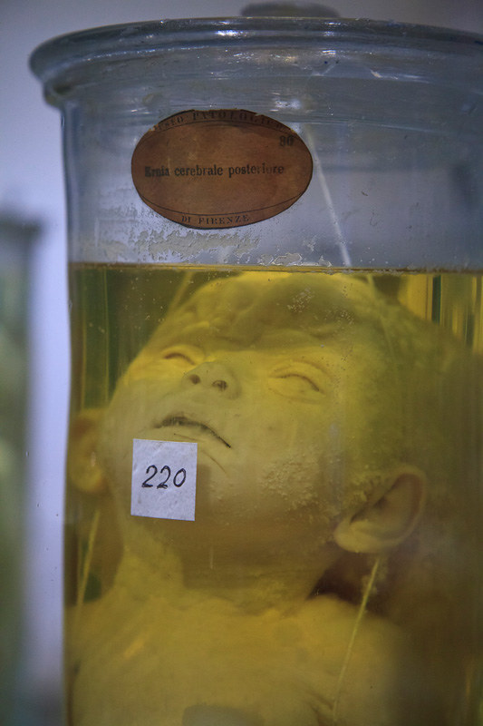
IMG_7394.jpgvon Morbid Anatomy
posted on Flickr:
Museo di Anatomia Patologica dell'Universitá degli Studi di Firenze; Full story on Morbid Anatomy here: bit.ly/WH5Cyh


Palate, complete* | October, 2015
Carbon dust rendering of palate and associated muscles.
*specimen was missing teeth.
Post link


Started this one out in a workshop with the mighty Kathryn Engberg, I added some extra sessions on my own and dropped a skeleton to study the pose. The triceps, teres major, teres minor and gluteus minimus are there just because I like them.
Post link

In honor of this, the week I am going to see Matt Nathanson in concert, I give you this infographic I made called Anatomy of a Girl in a Matt Nathanson Song. Tag yourself, I’m “the twist of your pigtails.”
Post link

Fig. 48. Digestive system of grain-eating birds. La vie : physiologie humaine appliquée à l'hygiène et à la médecine.1874.
Post link