#baby nursing student

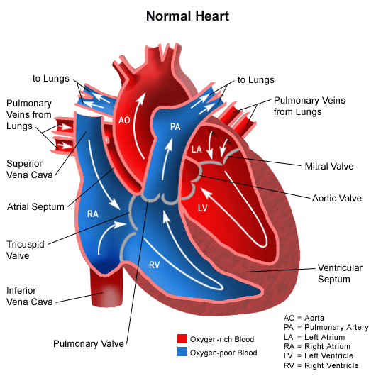
In order to better understand cardiac defects, it’s best to start off reviewing how a normal heart works. First, the Superior and Inferior Vena Cava carrydeoxygenated blood into the heart (Right Atrium) from other parts of the body. The deoxygenated blood then passes through the TricuspidValve into the Right Ventricle. Thedeoxygenated blood is following the pathway through the heart in order to get to the lungs to gain oxygen, next the blood passes through the PulmonaryValveand enters the Pulmonary Artery, the pulmonary artery is special because it is the only artery in the body that carries deoxygenated blood. Once the deoxygenated blood passes through to theLungsit becomes oxygenated. The newly oxygenated blood then flows back to the heart through the Pulmonary Veins and into the Left Atrium. It then passes through the MitralValveand into the Left Ventricle. The blood is then contracted through the AorticValveinto the Aortaand to the rest of the body.
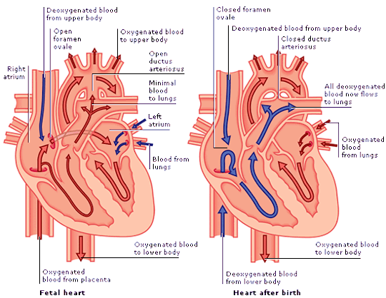
The Fetal Heart:
During the fetal period and some time after birth, the circulation is quite different. The heart has more, holes, if you will in order for the fetal blood to bypass the lungs that are unable to oxygenate blood while the fetus is in utero.
- The Foramen Ovale is an opening that allows passage of blood from the Right Atrium directly into the Left Atrium. The blood passing through is already oxygenated from the placenta.
- The Ductus Arteriosus is an opening that passes oxygenated blood from the Pulmonary Artery directly into the Aortato get pumped to the rest of the body.
Normal changes in the heart after birth:
- Ductus Arteriosus closes
- Foramen Ovale closes
- Ductus Venosus (connection from the umbilical cord) closes