#cardiac


My latest heart.
I know - another heart. I still love making them and, at this time of year, I get a lot of requests. Each is unique - sometimes on purpose, sometimes to cover up an accident - drill scratch, hole in the wrong place etc. The heart itself is vintage - new old stock from the 1970s - a lucite core with a thick copper plate. They are still available but, eventually, I’ll have to find an alternative. I’d love to find similar wood hearts - that could be hinged with a secret internal compartment. If anyone knows where something like that can be sourced - let me know. Dimensions would be 1 ½ inches wide/high and about ¾ of an inch deep so they could be cut and hollowed out.
The next one I’ll be making will be a bit different - a central “window” with a flashing red LED - with batteries that can be replaced. It will have to be a symmetrical design with a feature taking up that much real estate on the heart. I’m starting it tonight and I’ll post a video of it to show the “flash”.


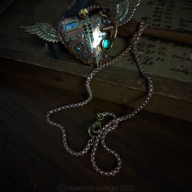
Silver wing heart - my latest winged mechanical heart with gears, gauges and glow in the dark grill. Lots of tiny details. Can be worn as a pendant, unique fob or just displayed. I can create a display box if required.
Post link


Framed Flying Heart
Here is one of my signature steampunk pieces - the mechanical flying heart. Although I make many of these, each one is unique with different features, layout and parts. I love working in miniature - the more complex - the better.
This piece is extremely intricate - 100+ parts some less than 1mm. The green glow under the grill/screen is from glow in the dark powder mixed with resin - I mix my own so it’s a strong glow. I recommend using a UV torch to “charge” it if wearing it at an event.
Nothing is glued on - it’s all deep pinned, soldered, using cold connection methods or screwed. It is robust enough the be worn without fear of anything falling off. I don’t cast or fabricate preferring to use existing items from a variety of sources. Some parts are antique, vintage, NOS or new.
Dimensions:
The pendant is 3 inches wide (75mm) and 1 ½ inches long (36mm) and 1 inch (24mm) deep. The chain is gold tone brass faceted cut cable link, 22 inches long with a gold tone lobster claw clasp.
The frame:
I decided to include a glass frame and dock when clients expressed their desire to be able to have the piece visible when not being worn. So I bought a commercial shadow box, painted it “distressed” gold, attached black felt and brass brackets. The heart can be accessed easily out of the back and attached to the chain with two push clasps and rings. The two clasp points keep the heart from flipping around when being worn.
It takes the piece to another level, especially if given as a gift. As a desk ornament or part of your home decor, my little biomechanical winged heart is sure to be commented on and desired.
The frame is square - 5 X 5 inches (125mm) and 1.6 inches (40mm) deep. The back panel is secured with four rotating clips and also has a “D” ring for wall hanging. The heart just rests on the two brass prongs.
Build time: 40 hours.
Available in my Etsy shop $455
Post link
In order to better understand cardiac defects, it’s best to start off reviewing how a normal heart works. First, the Superior and Inferior Vena Cava carrydeoxygenated blood into the heart (Right Atrium) from other parts of the body. The deoxygenated blood then passes through the TricuspidValve into the Right Ventricle. Thedeoxygenated blood is following the pathway through the heart in order to get to the lungs to gain oxygen, next the blood passes through the PulmonaryValveand enters the Pulmonary Artery, the pulmonary artery is special because it is the only artery in the body that carries deoxygenated blood. Once the deoxygenated blood passes through to theLungsit becomes oxygenated. The newly oxygenated blood then flows back to the heart through the Pulmonary Veins and into the Left Atrium. It then passes through the MitralValveand into the Left Ventricle. The blood is then contracted through the AorticValveinto the Aortaand to the rest of the body.
The Fetal Heart:
During the fetal period and some time after birth, the circulation is quite different. The heart has more, holes, if you will in order for the fetal blood to bypass the lungs that are unable to oxygenate blood while the fetus is in utero.
- The Foramen Ovale is an opening that allows passage of blood from the Right Atrium directly into the Left Atrium. The blood passing through is already oxygenated from the placenta.
- The Ductus Arteriosus is an opening that passes oxygenated blood from the Pulmonary Artery directly into the Aortato get pumped to the rest of the body.
Normal changes in the heart after birth:
- Ductus Arteriosus closes
- Foramen Ovale closes
- Ductus Venosus (connection from the umbilical cord) closes


chest pain yo; don’t forget these bad boys
click here to download all of my notes and study guides from medical school
Post link


Again, remember when the Tumblr hive mind was trying to convince people that Marvel was corporate propaganda because the anti-villain Cardiac was killing insurance people?
Yeah, it almost like those people either hadn’t read any of his appearances or took clipped panels out of context
Post link
Cardiology Echo Quiz/Case: Heart stops beating Part 1! SCD, VT, SVT, AVNRT?
Comment on this video on our Youtube-Channel: https://youtu.be/0BhAcmMroz0
Cardiology & Radiology Quiz/Case: Xray+Cardiac Devices. Can you spot them all? Pacemaker, ICD,…?
Watch and comment on Youtube: https://youtu.be/09Kp_wJvvWM

NURSE GANG. Where ever you are, I hope you are getting a chance to get outside and enjoy whatever weather you are having. Take some time to breathe some fresh air, close your eyes, and relax. Or go on a hike. Or lay in a pool. I chose to do all three. Here is to a being better and practicing self care!
open…
.
.
#selfcare #exceptionalnurses #poolday #aacn #nursegang #icunurse #intensivecare #criticalcare #ccunurse #icu #ccu #neuro #trauma #cardiac #nurse #nurses #nursing #nurselife #medsurg #nurseproblems #hospital #hospitallife #nursesofinstagram #nursesonly #nurseanesthetist #crna #nursememes #beard
Post link

Thanks for keeping it real, Boston. We didn’t get to see a whole heck of a lot of you but loved every bit we did get to see. The people of this city are truly the best.
.
.
.
.
.
#nti2018 #exceptionalnurses #ntiboston #aacn #nursegang #icunurse #intensivecare #criticalcare #ccunurse #icu #ccu #neuro #trauma #cardiac #nurse #nurses #nursing #nurselife #medsurg #nurseproblems #hospital #hospitallife #ricoh #cityskyline #skylines #longexposure #ricohgr #boston (at Boston, Massachusetts)
Post link

Nurses start conferences with mini raves.
.
.
.
.
#nti2018 #exceptionalnurses #ntiboston #aacn #nursegang #icunurse #intensivecare #criticalcare #ccunurse #icu #ccu #neuro #trauma #cardiac #nurse #nurses #nursing #nurselife #medsurg #nurseproblems #hospital #hospitallife #nursesofinstagram #nursesonly #nurseanesthetist #crna #nursememes (at Boston Convention & Exhibition Center)
Post link

Our super session today was led by @amycuddy and was all about being present for our patients and being confident. Amy talked about the use of open/expanded body positioning and it’s effects on confidence and feeling empowered. She has an awesome TED Talk on this subject. You should all check it out.
.
.
.
.
#nti2018 #exceptionalnurses #ntiboston #aacn #nursegang #icunurse #intensivecare #criticalcare #ccunurse #icu #ccu #neuro #trauma #cardiac #nurse #nurses #nursing #nurselife #medsurg #nurseproblems #hospital #hospitallife #nursesofinstagram #nursesonly #nurseanesthetist #crna #nursememes #powerposing #confidence #empowerment #ricohgr2 #ricohgr (at Boston Convention & Exhibition Center)
Post link

“As nurses we have to understand that our patients aren’t all going to fit in these neat little boxes that society has designed for them.”
I just had the opportunity to attend Jennifer Detchemendy’s session on Providing Safe, Sensitive Care to the LGBT(Q) Community. While I was aware of the issues the LGBT(Q) population encounters in regards to healthcare, I did not realize just how staggering the levels of discrimination were. A study in 2015 showed that 28% of transgender patients postponed seeking healthcare because of the fear of discrimination based on their previous healthcare encounters. 19% of those who did seek care reported being refused treatment because of their transgender or gender nonconforming status. In addition to fear of discrimination, seeking care is also delayed or difficult because of fears of breaches of confidentiality with HCPs, lack of partner benefits, prejudicial policies and procedures at both the organizational level and state level, a lack of LGBT health care providers, and being a subject of physical and/or verbal abuse by healthcare workers.
This should outrage all of you. Nurses need to be educated on these disparities, how to reduce these disparities, respecting the uniqueness of EVERY patient, and the importance of LGBT cultural humility. I appreciate Jennifer and NTI for getting the ball rolling on this education and pushing for creating a welcoming and judgment Free environment for our LGBT(Q) patient population.
.
.
.
.
.
#nti2018 #exceptionalnurses #ntiboston #aacn #nursegang #icunurse #intensivecare #criticalcare #ccunurse #icu #ccu #neuro #trauma #cardiac #nurse #nurses #nursing #nurselife #medsurg #nurseproblems #hospital #hospitallife #nursesofinstagram #nursesonly #nurseanesthetist #crna #nursememes #equality #humanrightscampaign #lgbt #lgbtq #equalrights #pride (at Boston Convention & Exhibition Center)
Post link
When you woke up at 5:30 to spend the whole day learning and you have one session left but none of the coffee spots in the convention center are open…
.
.
#nti2018 #exceptionalnurses #ntiboston #aacn #nursegang #icunurse #intensivecare #criticalcare #ccunurse #icu #ccu #neuro #trauma #cardiac #nurse #nurses #nursing #nurselife #medsurg #nurseproblems #hospital #hospitallife #nursesofinstagram #nursesonly #nurseanesthetist #crna #nursememes #powernap (at Boston Convention & Exhibition Center)

Time for the Super Sesh. AACN Presidential Address.
.
.
#nti2018 #exceptionalnurses #ntiboston #aacn #nursegang #icunurse #intensivecare #criticalcare #ccunurse #icu #ccu #neuro #trauma #cardiac #nurse #nurses #nursing #nurselife #medsurg #nurseproblems #hospital #hospitallife #nursesofinstagram #nursesonly #nurseanesthetist #crna #nursememes (at Boston Convention & Exhibition Center)
Post link

Tonight myself and some of my amazing peers got to represent Swedish American Hospital at the UW Organ and Tissue Donation Hospital Awards Celebration where we were presented with an Excellence in Tissue Donation Award. During the ceremony, we heard from the wife of a donor who discussed the positive impact donation and those caring for her husband made on her during such a tragic time. We also heard from a heart recipient about what her life was like while on the waiting list for her life-saving gift and the celebration that occurred when she was informed that they found a heart that was a perfect match. I am honored to be Co-chair of an Organ and Tissue Donation team within an organization that is full of nurses, RTs, physicians, and case workers who go above and beyond for our patients but especially for donors and donor families. We are constantly working to educate our community about the gift of donation and developing new tools to help donor families during such a difficult time. I love our partnership with UW Health and am am incredibly humbled by all of the work that is done by all of the UW OTD partner hospitals. #trustthebest
.
.
#organdonation #organandtissuedonation #uwotd #donatelife #nursegang #icunurse #intensivecare #criticalcare #ccunurse #icu #ccu #neuro #trauma #cardiac #nurse #nurses #nursing #nurselife #medsurg #hospital #hospitallife #nursesofinstagram #nursesonly #nurseanesthetist #opc #uwhealth #swedishamerican #swedishamericanhospital (at Monona Terrace Community and Convention Center)
Post link
There is quite a lot to be said about the medications we use for patients with arrhythmias. It’s easy to get lost as to what drugs do what and how, but thankfully there was a kind enough person by the name of Vaughan Williams, who actually broke them down into separate classes. Each class effects separate parts of the cardiac cycle, ultimately changing the electrical current of the heart.
Cardiac Action Potential
Before looking at the medications, we have to understand the cardiac cycle and how it actually works.

Source:x
The above chart presents the four phases of an action potential in a ventricular myocardial cell and how the electrolytes are used to cause the depolarization and repolarization of myocardial cells.
Phase 0 begins with a slight influx of sodium until it passes the potential threshold. Once past the threshold, more sodium channels will open and flood the cell, causing it the depolarize.
Phase 1 is an efflux of potassium from the cell, causing the cell to reach 0mV.
Phase 2 happens at this point. This is when calcium influx happens, prolonging the repolarization period. This period also goes by the name of an absolute refractory period for the cell, since it cannot depolarize during this time.
Phase 3 Calcium channels close again and potassium continues to efflux from the myocardial cell until the internal cell voltage returns to -90mV. Majority of potassium channels then close and the heart enters phase 4, which potassium is allowed to continue to leak into from the cell.
This process happens anywhere from 60 to 100 times per MINUTE!
Vaughan Williams Classifications
The major purpose of the medications in this class effect they way the cardiac action potential works in the cells of the heart. The drugs usually help to slow down specific phase to the heart and allow the heart to fix itself a bit.
Class I - Sodium Channel Blockers
These medications are designed to disrupt phase 0, causing a prolongation of it. There are 3 subcategories (a,b,c) that are broken down into moderate, weak, and strong.
This article won’t go into great depths, but the major goal of the class is to prolong the QRS complex and prolong or shorten QTi.
Medications include:
Lidocaine
Verapamil
Procainamide
Propafenone
Class II - Beta Blockers (-olol or -alol)
Quite commonly used out of hospital for patients with hypertension, beta blockers are actually a common antidysrhythmic. The basic pharmacology is: by blocking the beta-1 receptor sites, it prevents stimulation of the cardiac muscle to beat faster. The increase of sympathetic tone will decrease the rate the heart will beat.
Medications include:
- Propranolol
- Metoprolol
- Carvedilol
Class III - Potassium Channel Blockers
Similar to the Class Ia medications, potassium channel blockers are used to prolong APD, which can cause a prolongation of ERP. This class of medication is commonly known to treat different ventricular dysrhythmias (Vtach or Vfib). The most common medication for this class is Amiodarone and deserves a post of its own.
It’s most common use is during CPR, when the patient is in pulses Vtach or Vfib rhythm and is then followed by a drip with ROSC is achieved. An important note to make about Amiodarone is it can take 16 weeks to leave the system.
Medications include:
- Amiodarone
- Sotalol
- Ibutilide
Class IV - Calcium Channel Blockers
Commonly uses for Afib with RVR and PSVT, a calcium channel blocker will prolong phase 2 of the action potion in the cell. The goal is to slow the conduction through the atrioventricular (AV) node, slowing the ventricular tachycardia that is occurring. By prolonging the ERP in the AV node, the heart is able to regulate the rate better.
Calcium channel blockers are commonly prescribed by physicians to assist in the care of such arrhythmias. One side effect of these drugs is it may drop the patient’s BP, so ensure you have an SBP >100 or a MAP >65, prior to administering the medication
Class V - Others
This is the mix bag class. These drugs do not truly fit in any category but are still highly important to mention anyways. Two of these medications are Adenosine and Digoxin.
Adenosineprevents the re-entry of a signal in a sinus rhythm, preventing SVT. A warning though is for patients that have WPW syndrome may cause an increase in heart rate instead, so make sure you’re reading the rhythm correctly.
Digoxineffects vagal tone and is seen less as an emergency drug and more as a maintenance drug for chronic heart issues. A major issue with this drug is it holds a very narrow therapeutic index. Toxicity is quite possible if given too much.
Summary
Each class works in its own way on the action potential in the cardiac conduction system. How they affect the heart greatly determines when and how the medication should be used in different medical scenarios. The point of this article is to help a bit with the pharmacodynamics involved with the medications and to hint at the situations a person in the medical field would use them?
Remember to check out my facebook page. Feel free to send suggestions for possible article ideas, it might pop up some time. Always remember that medicine is an art, just as much as a science.