#medical school revision
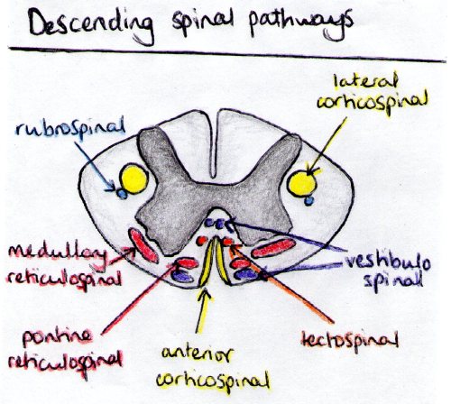
Descending tracts in the spinal cord. Descending tracts are generally motor, and are divided into pyramidal tracts (cotricospinal and corticobulbar - i.e. the voluntary ones) and extra-pyramidal (all the others).
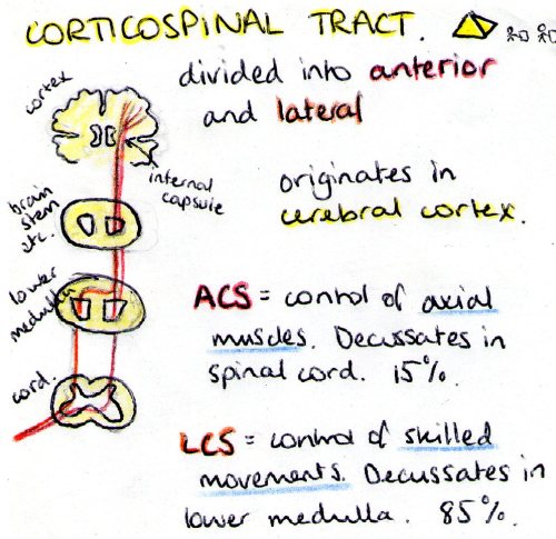
Corticospinal tract: carries motor fibres. Has an anterior (15%) and a lateral (85%) branch. Anterior controls the axial muscles while the lateral controlslimbs and skilled movements. They decussate in slightly different places, with the LCS crossing in the lower medullary pyramids, and the ACS at the spinal level they exit through.
Corticobulbar: not seen on this diagram because it doesn’t actually make it into the spinal cord. It terminates on cranial nerve motor nuclei to deal with motor functions of cranial nerves: facial expression, extra-ocular, etc.
Vestibulospinal: controls balance. It is special because it remains ipsilateral. Someone with a lesion of the vestibulospinal tract will fall (due to loss of balance control) towards the side of the lesion.
Reticulospinal: deals with reflexes. Has two branches, the pontine branch does extensor reflexes exclusively, while the medullary does both extensor and flexor. It also stays ipsilateral.
Tectospinal: another reflex type tract, that responds to visual and auditory stimuli. It is the reason that blind people can sometimes turn their head towards a flashing light without seeing it, but sensing it. Spooky.
Rubrospinal: bit vestigial in humans to be honest, most of its functions have been superseded by the corticospinal tract with which it joins in the lateral column of the spinal cord.
Post link
Support structures of the uterus. Weakening of any of these structures can lead to a uterovaginal prolapse.
Post link
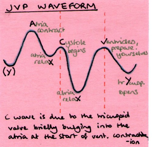
I can never remember what the different bits are on a JVP wave so here is a way to try and remember it. It’s a bit contrived, what with Cystole and trYcuspid but silly things are easier to remember.
C is the tricuspid valve closing and kind of bulging as it does. Imagine it over shooting ever so slightly then recoiling back into place.
V is the atria refilling, hence why the ventricles need to prepare for the next onslaught.
The letter refers to the bit of waveform that happened just before it. So when the atria contract they cause a bit of backflow, so the pressure in the jugular vein increases (there’s no valve into the atria, only out) causing the upswing. Then the atria relax so blood can flow into said atria, so the jugular vein empties and the pressure goes down.
Just remember that it’s the JUGULAR VENOUS pressure. Not the right atrial pressure. They’re similar, but not the same.
Post link
More detailed post-it of the corticospinal tracts, showing the two tracts and their decussation.
Post link


