#postmortem
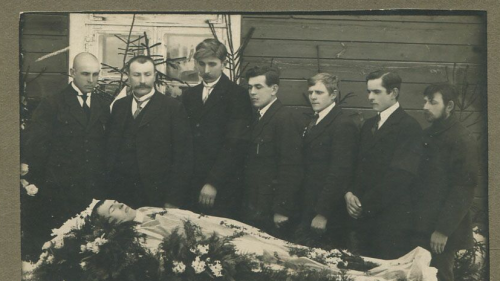
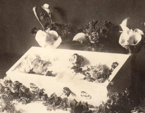
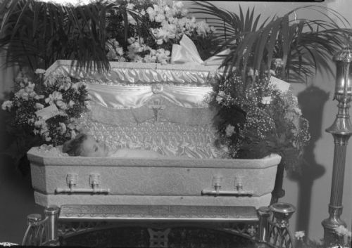
“William E. Crossen, eighteen-months-old, son of Mr. and Mrs. Phillip Crossen, shown in a coffin at Joyce Funeral Home.” Photo by Arthur M. Vinje. Madison, Wisconsin; July 26, 1945. Source: Wisconsin Historical Society.
Post link
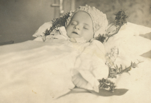
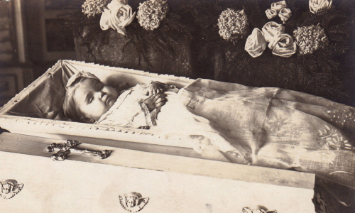
ca. 1880s-1910s, [hand tinted ambrotype, post mortem portrait of a child in a coffin, clutching a rosary and crucifix]
viaJeffrey Kraus Antique Photographics, Ambrotypes
Post link
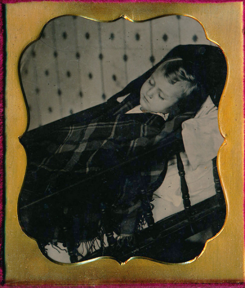
ca. 1860-80s, [hand tinted, postmortem tintype of a young woman]
viaJeffrey Kraus Antique Photographics, Tintype Collection
Post link
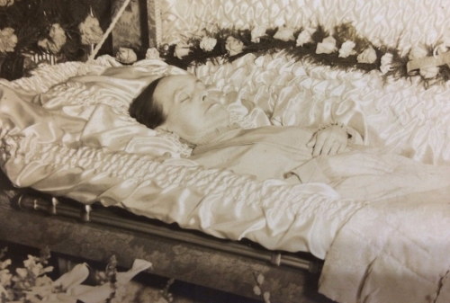
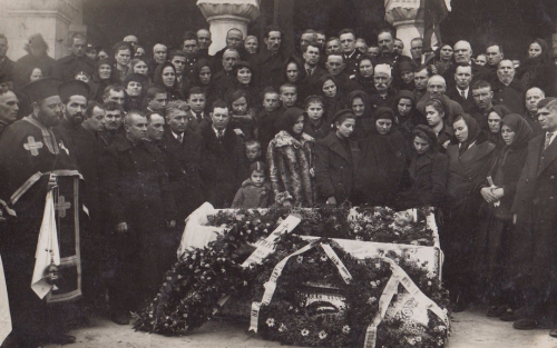
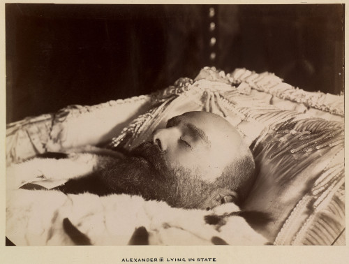
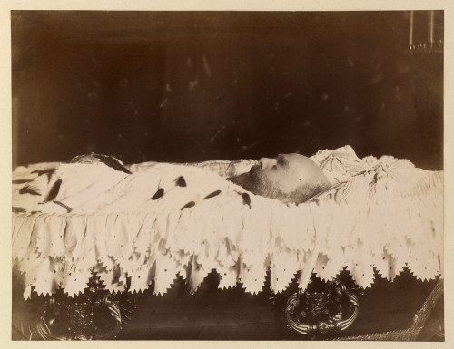
Emperor Alexander III Alexandrovich, “The Peacemaker” and “The Moujik Tsar”, lying in state, Livadia 1894
Post link
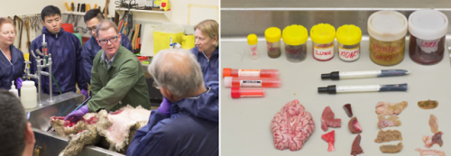
DPIRD Workshop
Last week I voluntarily attended a two day workshop at the Department of Primary Industries and Regional Development (aka the agricultural department). Unfortunately for me, the scheduled days alternated with my rostered night shifts at the equine hospital, meaning, aside from the odd power nap, I was awake from 9am on Monday to 9pm on Wednesday! That’s 60 hours of sleep deprivation that I brought on myself!
Despite having to physically hold my eyelids apart (because no amount of caffeine can substitute two nights of sleep), the workshop was well worth it. Only five students attended, so the training was interactive and hands on. Field vets, pathologists and other specialists contributed to the course, each imparting a wealth of knowledge and advice. A wide range of topics were covered over the two days, including:
- What to expect on our first farm visit
- Necropsies and sampling
- Disease surveillance and reportable diseases
- Foot and mouth disease (FMD)
- Transmissible spongiform encephalopathy (TSE)
- Avian necropsies and diseases
- Utilising the significant disease investigation (SDI) subsidy scheme
- Welfare
- Nutrition
We also had the opportunity to perform an entire sheep and chicken necropsy, including sample collection, independently. Necropsies are covered quite superficially by the vet course, and I previously had a limited understanding of the procedure. It was the first time I’d completed one by myself, and it was really helpful to receive direct feedback and advice from the experts. I also practiced removing the brain of the sheep with a hatchet and mallet (not to be confused with ‘mullet’). A field necropsy is something I could potentially be asked to do on my first day as a vet, so it’s great that I now have the confidence to ‘take the bull by the horns’, so to speak.
I found both days really interesting and beneficial to my first year out in the field! Everyone at the department was friendly and great to work with. It was clear that they were really invested in our education and more than willing to lend a hand or offer advice when needed.
Post link
Anatomical Pathology Rotation
I had low expectations for this rotation, after a friend described it as, “anatomical pathology…more like anatomical crapology”. Despite the negative review, I was determined to keep an open mind and start the week with fresh enthusiasm for dead things!
Monday began with a bit of pathology revision and a few practice cases. It was really hard and I felt as though I may as well have been listening to another language (situation not helped by the strong French accent of our teacher). In the afternoon, we got out the aprons, boots and safety glasses, and worked in groups to perform a couple of necropsies (the word for post-mortem examination in non-human animals). I worked on a five month old mixed breed dog that had presented to the hospital in acute respiratory distress. We systematically worked our way through all the anatomical structures, recording our findings as we went. While my teammates worked on other parts of the body, I removed the “pluck” - a weird term for a group of structures that can be removed together by cutting out the tongue and dissecting away attachments along the trachea and oesophagus to the heart and lungs. It is every bit as gruesome as it sounds! Pneumonia was evident in the lungs and the lung tissue was so dense it sunk when placed in water. I cut down the length of the trachea and discovered, to my surprise, the cause of death! A chunk of cartilage was wedged into the right primary bronchus, and had cut full thickness through the trachea. For one very brief moment I considered a career as a pathologist. I then looked around at all the death and gore surrounding me and quickly came to my senses. My friend, who was working on the gastrointestinal tract, found a huge number of nematodes (parasitic roundworms) in the intestines, which made my skin crawl!
On Tuesday we began the day with some more theory, and then headed to the post-mortem room in the afternoon for some more dead animals. A horse, a dolphin and a cat needed to be necropsied (no, this is not the start of a bad joke), and somehow I managed to get landed with the cat. Boo! It was a pretty straightforward case of chronic kidney disease, which meant I had time to hang around the dolphin and have a bit of a nosy. The dolphin, who was known to have a young calf, had died as a result of fishing line entanglement. The line had cut into her dorsal fin and tail fluke, restricting her movements and leaving open wounds susceptible to infection. It was a sad case and a reminder of the damage caused by human waste.
On Wednesday morning we went to the Department of Primary Industries and Regional Development (aka the agricultural department). We were given a tour of the facility, had a couple of lectures on necropsy sampling and notifiable diseases, and participated in a quick parasitology workshop. We headed back to uni in the afternoon for a cow necropsy. Large animal necropsies are quite a task, and it’s easy to become so focussed on what you’re doing that you don’t notice the absurdity of squatting in the middle of a cow carcass, soaked in blood, surrounded by organs and wielding a huge knife!
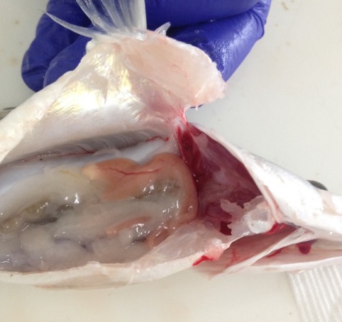
Thursday began with some more practice cases. Our teacher broke the news to us that necrosis is in fact white, rather than black as we had all previously been led to believe. Mind blown! In the afternoon we went to the university fish health unit where we anaesthetised fish and practiced taking gill samples and skin scrapes. We then euthanised the fish with an anaesthesia overdose, followed by cutting the gill arches and severing the spinal cord, just to be sure. Once dead, we performed a necropsy and collected samples for a research project. The tiny heart continued to beat for many minutes after being removed from the body. Creepy-deepy!
The final day of the rotation involved assessed pathology rounds and an online case-based exam. For rounds we had to present a specimen collected during the week to our peers and a small audience of vets from the teaching hospital. I presented the trachea and lungs from the case on Monday. I was really nervous but it actually went much better than I expected. I even survived the brutal questioning from the pathologists and got complimented on my explanation of the pathophysiology of pneumonia! The exam, on the other hand, was straight out of hell. The questions were so random and specific - there was no way we could’ve known the answers! Luckily we we were all in the same boat. All in all, anatomical pathology is a great rotation for those who enjoy feeling like an idiot constantly. Although I do enjoy the odd necropsy, I think it’s fair to say a career in pathology is not for me!
Post link


![ca. 1860-80s, [hand tinted, postmortem tintype of a young woman]via Jeffrey Kraus Antique Photograph ca. 1860-80s, [hand tinted, postmortem tintype of a young woman]via Jeffrey Kraus Antique Photograph](https://64.media.tumblr.com/43ad5904049b45ae8ce0330611ad8a62/tumblr_nph87uMNu11qa51rdo1_500.jpg)

