#zebrafish

24 hours in the life of a zebrafish embryo
In this video you can watch 24 hours (the second day) in the development of a tiny zebrafish embryo.
Some cells have been labelled with a fluorescent protein naturally found in jellyfish. This fluorescent label marks several developing organ systems including the eye, hatching gland and kidney.
You can also see two groups of cells migrating along the trunk of the embryo, depositing smaller clumps of sensory cells as they go. These cells are the precursors of the lateral line system, which gives fish a sense of ‘touch at a distance’, enabling them to shoal, avoid obstacles and find prey.
Look more closely to see individual cells moving around over the skin.
Light sheet microscopy illuminates the specimen with a thin sheet of light and allows scientists like Dr Tanya Whitfield and her team at the University of Sheffield to observe cells and embryos at high speeds or for long times. This research is helping them to gain a better understanding of the amazing process of embryogenesis, including the development of sensory systems such as the lateral line and inner ear.
Video: Sarah Baxendale and Nick van Hateren.
See more beautiful images of zebrafish embryos here


A two-day-old zebrafish embryo brain
These striking images show a zebrafish embryo brain at just two-days-old as seen with a Light Sheet Fluorescent Microscope.
Light sheet microscopy illuminates the specimen with a thin sheet of light and can be used to see relatively large structures in fine detail. You can see all the nerve cells and connections in the zebrafish brain stained with an antibody in red.
Being able to see embryos in this detail allows research teams at the University of Sheffield to follow the amazing process of embryogenesis, from a single cell (the fertilised egg) to a functioning animal with hundreds of different cell types.
Images: Sarah Baxendale, Stone Elworthy and Nick van Hateren.
Post link


Estrogens alleviate hyperactivity in ‘autistic’ zebrafish
The female sex hormone estrogen reduces sleep disruption in zebrafish genetically designed to help understand autism spectrum disorder (ASD) scientists have discovered.
Researchers from the University College London In collaboration with scientists at Yale University and University of California, San Francisco, were investigating the function of genes linked to autism and seizures in humans by using zebrafish as a model system. They unexpectedly discovered that estrogens calm hyperactive fish during the night, which will help scientists to understand the brain pathways affected in ASD.
The finding is intriguing given ASD is four times more common in men than women. All humans produce estrogen, but levels are significantly higher in women than men.
These images show zebrafish brains, with axon tracts, neurons and interneurons tagged in different colours.
This research helps scientists to understand the function of an autism risk gene in the developing brain, which is important for understanding the biology of autism.
Images: Kate Turner, UCL
Post link
In really surreal science news of the day, researchers have now found a way to watch how hundreds of individual cells work together to maintain and regenerate skin tissue, thanks to a genetically engineered line of technicolor zebrafish.
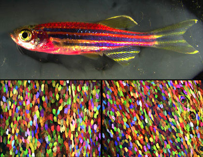
(To the naked eye, this genetically engineered zebrafish has a magenta tone (top). But under a microscope, every cell has a distinct color, thanks to a new labeling technique called Skinbow (bottom). Photo credit: Chen-Hui Chen, Duke University)
Ken Poss, a professor of cell biology at Duke University, and his lab have spent the last few years creating “Skinbow,” a mutant zebrafish with fluorescent markers embedded in the DNA code of its skin cells. Their study was published on March 21st, 2016 in Developmental Cell, and is available for free download: Multicolor Cell Barcoding Technology for Long-Term Surveillance of Epithelial Regeneration in Zebrafish.
Each of Skinbow’s color markers can be expressed in three different ways, resulting in red, green, or blue fluorescence. With three color options and roughly a hundred copies of the gene per cell, the total number of colors any cell can take on is probably in the thousands.
The research team has now been able to observe tissue regeneration by scraping a few cells off the fish’s fin, and visualize the healing process on a cellular level. Any color patterns that develop during tissue regeneration are inferred to arise from a single cell.
“It is like you have given each cell an individual barcode,”said Chen-Hui Chen, a postdoctoral fellow in Poss’s lab and lead author on the study. “You can precisely see how individual cells collectively behave during regeneration.”
Listen to Ken Poss discuss the experiment:
“One thing we weren’t expecting is that within a few hours of injury, cells that are spared acquire some mobility on the surface,”Posssaid in an interview with Gizmodo. “They also expand in size, some doubling. Then there’s a quick wave of replacement—you can see new cells emerging from the layer underneath.”
What is even more fascinating is that cells behaved differently in function of the injury. For example, following fin amputation, pre-existing cells were initially recruited from below the amputation plane to cover the wound. Then, new cells were generated at a rapid pace, and in the end some cells at the tip of the regrown tissue temporarily grew in size to provide surface coverage.

(Here, the multicolor cells are evident on the exposed portion of a dissected scale. Photo credit: Chen-Hui Chen, Duke University)
Poss is hopeful that the Skinbow technique can be extended to other species in the future, even humans. It obviously would involve getting the right set of genetic tools, and developing a way to image tissue continually over time, but Poss is hopeful that doctors may be able to use this technology to visualize how human tissue responds to new cancer drugs, or treatments meant to accelerate healing.