#neurons
A two-day-old zebrafish embryo brain
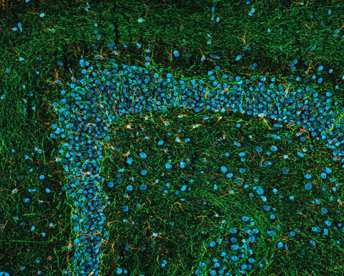
These striking images show a zebrafish embryo brain at just two-days-old as seen with a Light Sheet Fluorescent Microscope.
Light sheet microscopy illuminates the specimen with a thin sheet of light and can be used to see relatively large structures in fine detail. You can see all the nerve cells and connections in the zebrafish brain stained with an antibody in red.
Being able to see embryos in this detail allows research teams at the University of Sheffield to follow the amazing process of embryogenesis, from a single cell (the fertilised egg) to a functioning animal with hundreds of different cell types.
Images: Sarah Baxendale, Stone Elworthy and Nick van Hateren.
Post link
We tend to think our memory works like a filing cabinet. We experience an event, generate a memory and then file it away for later use. However, according to medical research, the basic mechanisms behind memory are much more dynamic. In fact, making memories is similar to plugging your laptop into an Ethernet cable—the strength of the network determines how the event is translated within your brain.
Neurons (nerve cells in the brain) communicate through synaptic connections (structures that pass a signal from neuron-to-neuron) that “talk” to each other when certain neurotransmitters (chemicals that allow the transmission of these signals) are present.
Think of a neurotransmitter as an email. If you’re busy and you receive one or two emails, you might ignore them. But, if you are bombarded with hundreds of emails from the same person, saying basically the same thing, all at the same time, you will likely begin to pay attention and start a conversation with the sender: Why on earth are you sending me all these emails?
Similarly, neurons only open a line of communication with each other when they receive stimulation from several of the same neurotransmitters at once: Oh, my neighbor keeps hitting me with the same signal? I better talk to them! So, how exactly does this relate to memory? It’s the strength of these connections between neurons that determines how a memory is formed.
“The persistent strengthening of these activated synapses (connections) between neurons is called long-term potentiation (LTP),” said William Griffith, Ph.D., a cellular neuroscientist and chair of the Department of Neuroscience and Experimental Therapeutics at the Texas A&M Health Science Center College of Medicine. “LTP is the most recognized cellular mechanism to explain memory because it can alter the strength between brain cell connections. If this strength is maintained, a memory can be formed.”
LTP happens when nerve cells “fire” or talk to one another at an elevated rate without further increased stimulation from neurotransmitters. In a sense, it’s like building a relationship with the email sender. Once you’ve started a dialogue with the sender you’re in a better position to communicate more easily and maintain a strong rapport. Just like you might add the sender to your contact list, your brain has created a ‘strengthened synaptic contact.’ But, if you’re not talking, the relationship wanes.
Likewise, your ability to recall and remember certain memories depends on maintaining the strength of this long-term connection between synaptic contacts. LTP acts as an Ethernet cable of sorts—allowing your brain to upload, download and process at a higher rate—which may explain why some memories are more vivid than others: the pathway on which you contact them performs at a faster pace.
“The brain is a plastic organ,” Griffith explained. “This means it can easily reconfigure or modify itself. However, it’s also a muscle. You use it or you lose it. As the synapses and pathways between neurons are used, they gain the ability to become strengthened or permanently enhanced. This is the building block of how memory works.”
In the same vein, losing this strong LTP— or heightened synaptic connections between neurons—could be the reason behind cognitive loss and impairment. “Because the brain is an organ, it will show wear and tear,” Griffith continued. “Many people believe this decrease in neurons ‘talking’ to one another is responsible for cognitive loss—because the pathways are not being used or strengthened. Just as muscles in the body atrophy when you don’t use them, the brain will deteriorate when it’s not stimulated.”
Griffith said the argument about how memory is consolidated and retrieved is vast, and there are many aspects that still need to be studied about the phenomenon. “When you look at or smell something, it contributes to your memory of an event,” he said. “This can be mapped in many parts of the brain. Memory may also be involved in certain behaviors like addiction. Why does this happen? Is it because the pathways for addiction are strengthened, or because they’re repressed? We don’t know yet.”
The science behind memory is a complex one, and will likely be studied for decades to come. “Many different pathways in the brain interact to set up complex circuits for different types of memories,” Griffith said. “There’s much debate and more research that needs to be done to fully comprehend how our brain generates, consolidates and retrieves memories.”
Post link
Study reveals how differences in male and female brains emerge
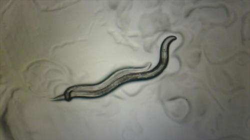
Nematode worms may not be from Mars or Venus, but they do have sex-specific circuits in their brains that cause the males and females to act differently. According to new research published in Nature, scientists have determined how these sexually dimorphic (occurring in either males or females) connections arise in the worm nervous system. The research was funded by the NIH’s National Institute of Neurological Disorders and Stroke (NINDS).
“For decades, there has been little focus on the impact of sex on many areas of biomedical research,” said Coryse St. Hillaire-Clarke, Ph.D., program officer on this NINDS project. “This study helps us understand how sex can influence brain connectivity.”
In nematode worms, (known as Caenorhabditis elegans or C. elegans), a small number of neurons are found exclusively in male or female brains. The remaining neurons are found in both sexes, although their connection patterns are different in male and female brains. Oliver Hobert, Ph.D., professor of biological sciences at Columbia University in New York City, and his colleagues looked at how these wiring patterns form.
Dr. Hobert’s team observed that in the worms’ juvenile state, before they reach sexual maturity, their brain connections were in a hybrid, or mixed state, comprised of both male and female arrangements. As they reached sexual maturity, however, their brains underwent a pruning process, which got rid of particular connections and led to either male or female patterns.
“We found that differences in male and female brains develop from a ground state, which contains features of both sexes. From this developmental state, distinctly male or female features eventually emerge,” said Dr. Hobert.
Next, Dr. Hobert’s team showed that sex-specific wiring in the brain results in dimorphic behavior. They discovered that PHB neurons, chemosensory brain cells that detect chemical cues in the environment such as food, predators or potential mates, work differently in males and females. In males, these neurons proved to be important in recognizing mating cues while in females, the neurons helped them avoid specific taste cues. However, early in development, PHB neurons in males also responded to signals regulating taste, suggesting that even though those neurons are found in all nematodes, in adults, their functions differ as a result of sex-specific wiring in the brain.
Dr. Hobert’s team used genetically engineered nematodes to look more carefully at individual connections between brain cells. The researchers found that swapping the sex of individual neurons changed wiring patterns and influenced behavioral differences in males and females.
Additional experiments helped to identify genes involved in regulating the pruning process during development. Dr. Hobert’s group discovered that certain transcription factors, which are molecules that help control gene activity, are present in a dimorphic state and may help establish male or female connections in the brain. In future experiments, Dr. Hobert and his colleagues plan to examine how these molecules target specific connections for pruning.
Post link
Splashing neurons with colour to track their development.
A big problem when trying to monitor the ways in which cells change over time is that of consistently identifying the same one. In the tangled billions of neurons in animal brains, this careful art only becomes more complicated.
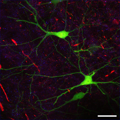
A research team headed by the University of Southampton has described a possible solution to this problem: use a viral vector to uniquely colour individual cells to keep track of them. Pretty much any RGB colour can be chosen to paint cells as is needed. This way, the problem is reduced to a matter of finding the ‘red one’ among the rest.
The colouration of cells marked in this way is permanently ingrained in DNA, and so is inherited by any daughter cells produced. Using this technique, scientists may soon better understand how connections are made in the brain, how early embryonic development occurs and more simply by watching those processes at the cellular level.
Post link

A recent paper in the journal Neuron presents a discovery of a bi-directional neural network connecting the legs and the

lil neuron stick n poke \ʕ •ᴥ•ʔ/ offering free stick n pokes on simple designs in london!!
Clashing synapses
.
.
.
#blackandgrey #blackandwhite #bwphotography #abstractart #abstract #trippyartwork #trippy #neurons #synapses #tree #naturephotography #natureatitsbest #collageart #collage #photography #opart #opticalart #monochrome #perfectnature #repetition #repeatingnature
https://www.instagram.com/p/B0bI3W1iqN2/?igshid=1o9nv4fz80ty7
Post link
While 28 weeks pregnant you’ve reached the third trimester in your pregnancy. Your baby’s also developing billions of neurons in it’s brain, and adding more body fat in preparation for life in the outside world. http://bit.ly/128CiS9
Post link
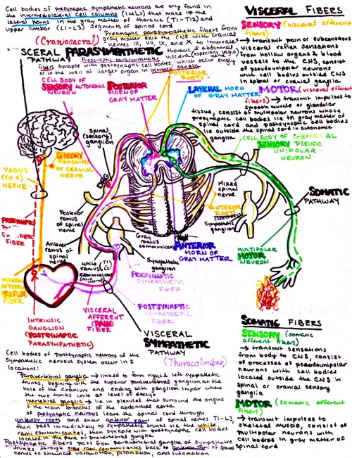
Autonomics and their trajectory in the spinal cord
download my medical school and anatomy notes & study guides here!
Post link
ALS-linked gene found switched on in new bits of the brain
ALS, also known as Motor Neuron Disease, is a fatal neurodegenerative condition for which we don’t have a cure and don’t fully understand.
We do know that a gene called C9orf72 is often involved, and many patients have abnormal repeated sequences within the gene causing neurons in the brain to die. The gene is also implicated in a type of dementia called Frontotemporal Dementia (FTD).
Now scientists studying it in mouse brains have found it is switched on in two regions that we didn’t know about before.
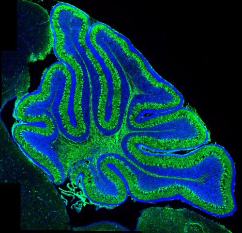
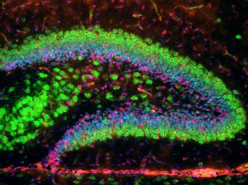
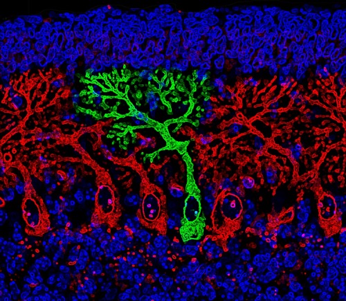
They discovered that C9orf72is strongly expressed in the hippocampus - containing adult stem cells and which is important for memory - and the olfactory bulb - which is involved in the sense of smell.
Loss of smell is sometimes a symptom in FTD.
The University of Bath team hope the findings will help researchers gain a better understanding of C9orf72 ′s role in both diseases and help map where it is switched on and off as the brain develops.
This could help us figure out new ways to slow down, treat or even cure the symptoms and explain why people born with abnormalities in C9orf72don’t develop symptoms until decades after birth.
Images: Andrew L Bashford and Vasanta Subramanian
The top and bottom images show mouse cerebellum stained to reveal the Purkinje neurons (green).
The middle image shows the dentate gyrus of the mouse hippocampal formation, which contributes to the formation of new memories stained for neurons (green) and stem cells (red).
Post link


