#pharmacology

Tea Time ☕️

Pharmacology ☕️


Really nice morning.

What a nice evening ☕️

☕️ cute afternoon, isn’t it ?

☕️


Happy New Year

Pharmacology ☕️

Pharmacology ☕️
Thalidomide was originally introduced as a non-barbiturate sedative in 1957 and later marketed for the treatment of nausea in pregnant women by the German company Chemie Grünenthal, as antiemetic properties were discovered. In the 1960s however it became apparent that thalidomide treatment resulted in severe teratogenic birth defects and from November 1961 it began to be withdrawn worldwide. It has since been reintroduced into a number of studies and treatments due to its immunosuppresive and anti-angiogenic activity.

Chemical structure of thalidomide. Thalidomide is a stereo-isomer and can exist, depending on the state of the chiral carbon, in two enantiomeric states. Both R and S can interconvert in body fluids and tissues. Thalidomide was distributed as a racemic mix of both enantiomers.
- Thalidomide is a piperidinyl isoindole and synthetic derivative of glutamic acid, consisting of two linked glutarimide and pthalimide rings.
- The unstable chiral carbon allows two enantiomers to coexist - the S-enantiomer being teratogenic (Franks, 2004.)
- The mechanism of action that resulted in these teratogenic effects is still not fully understood.
- Leading theories focus on thalidomide’s antiangiogenic properties; ability to induce cell death and generate reactive oxygen species; and Cereblon, a thalidomide-binding protein and primary source of teratogenic action (Takumi, 2010).
- Thalidomide is hydrolyzed in bodily fluids and metabolized in the liver by cytochrome p450.
When first developed thalidomide was deemed so non-toxic that an LD50 could not be established, but tragically an estimated 25,000 babies were born severely impaired and a further 123,000 miscarried or stillborn (Johnson, 2016).

Antiangiogenic properties
Thalidomide has the ability to inhibit angiogenic vascularization in embryos, thus carrying out teratogenic damage.
- The bulk of its damage occurs in days 20-36 of fertilization.
- During this time, the primitive vessels formed by vasculoneogensis mature and begin to proliferate and migrate in response to signals and growth factors such as FGF.
- It is thought that analogues of thalidomide could block a number of these signals, thus preventing embryonic vascular development outwards towards the limbs and resulting in limb deformities such as phocemelia.
Molecular targets of thalidomide
Cereblon
- Forms part of a ubiquitination complex with DNA Binding Protein 1, which in turn selects molecules for destruction.
- Thalidomide binds, preventing formation of the complex and proper regulation of developmental signaling molecules, thus initiating teratogenis by inhibiting angiogenesis (Takumi, 2010).
- Could also degrade some proteins and prevent breakdown of others.
Tubulin
- part of the cytoskeleton and required for cell proliferation and formation of new vessels in the embryo.
- When bound by the 5HPP-33 thalidomide analogue, cytoskeletal dynamics are altered preventing cell division.
- This could prevent cell proliferation and migration and consequently tissue morphogenesis, the severity of which would be dependent on vascular and tissue maturation at the point of contact with thalidomide (Vergesson, 2015).
Amphetamines are sympathomimetic amines which mimic the structures of the catecholamine neurotransmitters, noradrenaline and dopamine.
- They are substrates for the neuronal plasma membrane monamine uptake transporters DAT and NET (dopamine and norepinephrine transporters)
- Thus act as competitive inhibitors, reducing the reuptake of dopamine and noradrenaline.
- In addition, they enter nerve terminals via the uptake processes or by diffusion and interact with the vesicular monoamine pump VMAT-2 to inhibit the uptake into synaptic vesicles of cytoplasmic dopamine and noradrenaline.
- The amphetamines are taken up into the storage vesicles by VMAT-2 and displace the endogenous monamines from the vesicles into the cytoplasm.
- At high concentrations amphetamines can inhibit monoamine oxidase, which otherwise would break down cytoplasmic monamines.

The cytoplasmic monoamines can then be transported out of the nerve endings via DAT and NET transporters working in reverse, a process thought to be facilitated by amphetamine binding to the transporters. The concentration of extracellular dopamine and noradrenaline is thereforeincreased, and the reward response generated by increased activation of dopamine receptors can eventually lead to addiction.
The biggest problem facing addiction is the lack of effective medical treatment available. Currently, therapeutic strategies involve giving the patient something similar, but less potent than the drug of addiction, in an attempt to slowly reach abstinence with minimal withdrawal. However, due to memory and learning mechanisms as well as epigenetic and transcriptional changes, relapse is not uncommon.
Pharmacology is less scary than it looks but hard to understand just through reading - here’s a great video
The human reward system is made up of neural structures responsible for incentive salience (desire), associative learning (primarily positive reinforcement and classical conditioning), and positive/pleasure emotions (e.g. euphoria).
- Dopamine is the primary neurotransmitter of the brain’s reward mechanisms
- Most important reward pathway is the mesolimbic dopamine pathway.
- Thisconnects the ventral tegmental area (VTA) of the midbrain, to the nucleus accumbens (NAc) and olfactory tubercle, which are located in the ventral striatum
- Theprojections from the VTA are a network of dopaminergic neurons with co-localized postsynaptic glutamate receptors.
- The NA itself consists mainly of GABAergic medium spiny neurons.
When a rewarding stimulus, such as eating food, or direct stimulation by a drug occurs, dopaminergic neurons in the VTA are activated. These neurons project to the NAc, and their activation causes dopamine levels in the NAc to rise,activating dopamine receptors and generating a reward response, thus encouraging repetition and learning. –> any activity that resulted in a reward from your brain will therefore be one you want to repeat. This is essentially how your brain keeps you alive and reproducing.

Another major dopamine pathway, the mesocortical pathway, also originates in the VTA but travels to the prefrontal cortex, and is thought to integrate information which determines whether a behavior will be elicited.Thebasolateral amygdala projects into the NAc and is thought to also be important for motivation, while the hippocampus plays a role in learning and memory.
Even though increased dopamine in the brain reward system is generally thought to be the final common pathway for the reinforcing properties of drugs, other neurotransmitters such as serotoninare involved in the modulation of both drug self-administration and dopamine levels. Serotonin may be important in modulating motivational factors, or the amount of work and individual is willing to perform to obtain a drug. Serotonergic neurons project both to the NA and VTA and appear to regulate dopamine release at the NA.
- Excessive intake of addictive drugs –> repeated release of high amounts of dopamine –>increased dopamine receptor activation.
- The intrinsic purpose of an endogenous reward center is to reinforce behaviors that promote survival, so when a drug stimulates this center, drug-seeking behavior is also promoted - induced by glutamatergic projections from the prefrontal cortex to the nucleus accumbens
- Prolonged and abnormally high levels of dopamine in the synaptic cleft can induce receptor downregulation, resulting in a decrease in the sensitivity to natural stimuli.
- Alongside the positive reinforcement, these withdrawal symptoms can be considered negative reinforcing factors.
- Discontinued drug use will often induce various negative responses such as chronic irritability, physical pain, emotional pain, malaise, dysphoria, alexithymia, and loss of motivation for natural rewards.
Chronic addictive drug use causes alterations in gene expression in the mesocorticolimbic projection, which arise through transcriptional and epigenetic mechanisms. The most important transcription factors that produce these alterations are ΔFosB, cyclic adenosine monophosphate (cAMP), (CREB), (NF-κB). Overexpression of ΔFosB in the D1-type medium spiny neurons in the nucleus accumbens is necessary and sufficient for many of the neural adaptations and behavioral effects (e.g., expression-dependent increases in drug self-administration and reward sensitization) seen in drug addiction. This means an individuals actual genes are changed by chronic drug use to make them even more addicted - not just a case of being able to stop when they chose.
Normal metabolism
- Drug metabolizing enzymes are are located in lipophilic membranes of endoplasmic reticulum (Cytochrome P450 (2E1, 1A2 and 3A4))
- 95% of paracetamol is conjugated with glucaronic acid or sulphate and excreted in urine. This is non-toxic.
- 5% is oxidized by the CYPs to form N-acetyl-P-benzoquinone imine (NAPQI), which is toxic.
- NAPQI is conjugated with glutathione and mercapturic acid and cysteine conjugates and excreted in bile in normal metabolism.

In overdose
- the glucaronic acid and sulphate pathways become saturated, so more goes via the NAPQI pathway.
- Glutathione stores become depleted as it is consumed by the NAPQI.
- NAPQI accumulates and begins conjugating hepatic proteins and nucleic acids, including those of the mitochondria.
- Enzymes such as glutathione peroxidase, HMG-CoA, become inhibitedandradicals begin forming as the mitochondria are damaged.
- In the cytosol, MAP kinase JNK is activated, which binds to Sab proteins on the outer mitochondrial membrane, which in turn deactivates p-Src on the inner mitochondrial membrane.
- This inhibits electron transport.
- Moreradicalsare released and there is more oxidative stress.
- There is mitochondrial and cell death leading to necrosis if not treated early enough.
Liver function tests:
- GGT elevated
- Bilirubin elevated
- ALP elevated
- AST and ALT extremely high
- Becoming higher than 100 times the upper reference limit in toxic ingestion. No other hepatic conditions increase AST/ALT to these levels.
- Decreased albumin once necrosis forms – many other conditions of the liver don’t reach this stage
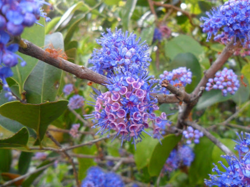
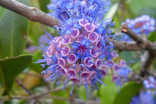
Ironwood tree - Memecylon umbellatum
Also referred to as Delek Air tree, Memecylon umbellatum (Myrtales - Melastomataceae), is a large shrub or small tree, up to 8-14m tall, that produces showy clusters of amazing bright purple flowers, about 1 cm each.
This tree, which can be found in Peninsular India and Sri Lanka, is not only beautiful, but also useful since it has several traditional and pharmacological uses. It provides hard timber used for building houses and boats, and a yellow dye can be extracted from the leaves and the bark that is used to treat bruises and various ailments.
Photo credit: Photo credit: ©Nuwan Chathuranga | Locality: Sri Lanka (2014)
Post link
Thanks for a great class today! Bought some new apothecary jars for in the shop from @alchemy_and_ashes
#apothecary #toxicology #pharmacology #poisonhistory #toxicology #thepoisonaffairs #ethnobotany #poisonpath #thepoisonpath #poisonmagic #poisonousplants #poisonlore #deadlyplants #poisonbooks #toxicologybooks #booksonpoison #thepoisonpath #veneficium # (at Alchemy & Ashes)
https://www.instagram.com/p/CdRRHs1re3P/?igshid=NGJjMDIxMWI=
Post link


