“All the eggs a woman will ever carry form in her ovaries when she is a four month old fetus in the womb of her mother. This means our cellular life as an egg begins in the womb of our grandmother. Each of us spent 5 months in our grandmother’s womb. And she in turn formed within the womb of her grandmother. We vibrate to the rhythms of our mother’s blood before she herself is born.”
Demons probably get some things with humans confused with animals I think.
- Mammon freaking out at Beel sharing a snack with MC. Not because he wanted them apart, but because “BEEL NO HUMANS CANT EAT GRAPES”, followed by a forceful full body squeeze Heimlich maneuver. (He was thinking of dogs.)
- It’s MC and Belphies night to cook dinner. A particularly difficult to open lid results in MC gets salt all over themselves. Belphie, half asleep at the table JUMPS UP and frantically wipes the human off/shakes them so the salt goes flying everywhere. He grabs MC by the shoulders and observes their skin closely for any signs of burns/melting. There are tears in his eyes. (He was thinking of slugs.)
- Asmos closet is a mess. He’s struggling to pair an outfit together with a somewhat limited color palette so that MC and Solomon could appreciate his beauty, even with their sad, pathetic, limited color perception :(
Tonsillitis is inflammation of the pharyngeal tonsils.
Inflammationusually extends to the adenoid and the lingual tonsils
Most commonly viral
Most cases of bacterial tonsillitis are caused by group A beta-hemolytic Streptococcus pyogenes (GABHS) - strep throat.
Spread through the air.
Symptoms may include sore throat, fever, enlargement of the tonsils, trouble swallowing, and large lymph nodes around the neck. Complications include peritonsillar abscess.
Recurrent tonsillitis
A polymicrobial flora consisting of both aerobic and anaerobic bacteria
Other competing bacteria are reduced - less interference to GABHS infection.
Streptococcus pneumoniae, Staphylococcus aureus, and Haemophilus influenzae are the most common bacteria isolated in recurrent tonsillitis, and Bacteroides fragilis is the most common anaerobic bacterium isolated in recurrent tonsillitis.
The microbiologies of recurrent tonsillitis in children and adults are different; adults show more bacterial isolates, with a higher recovery rate of Prevotella species, Porphyromonas species, and B fragilis organisms , whereas children show more GABHS. Also, adults more often have bacteria that produce beta-lactamase.
Chronic tonsillitis
Polymicrobial bacterial population present
There is likely a relationship between tonsillar size and chronic bacterial tonsillitis based on both the aerobic bacterial load and the absolute number of B and T lymphocytes.
Fewer dendritic cells on the surface epithelium and more in the crypts and extrafollicular areas during chronic tonsillitis.
Radiation exposure may relate to the development of chronic tonsillitis. A high prevalence of chronic tonsillitis was noted following the Chernobyl nuclear reactor accident in the former Soviet Union.
The tonsils are a set of lymphoid organs consisting of the adenoid tonsil, two tubal tonsils, two palatine tonsils, and the lingual tonsils. These organs play an important role in the immune system.
Palatine tonsil
Main tonsils
Non-keratinized stratified squamous epithelium
Incompletely encapsulated
Long, branched crypts
Located on sides of oropharynx between palatoglossal and palatopharyngeal arches
Reach their largest size in puberty, and they gradually undergo atrophy
Thalidomide was originally introduced as a non-barbiturate sedative in 1957 and later marketed for the treatment of nausea in pregnant women by the German company Chemie Grünenthal, as antiemetic properties were discovered. In the 1960s however it became apparent that thalidomide treatment resulted in severe teratogenic birth defects and from November 1961 it began to be withdrawn worldwide. It has since been reintroduced into a number of studies and treatments due to its immunosuppresive and anti-angiogenic activity.
Chemical structure of thalidomide. Thalidomide is a stereo-isomer and can exist, depending on the state of the chiral carbon, in two enantiomeric states. Both R and S can interconvert in body fluids and tissues. Thalidomide was distributed as a racemic mix of both enantiomers.
Thalidomide is a piperidinyl isoindole and synthetic derivative of glutamic acid, consisting of two linked glutarimide and pthalimide rings.
The unstable chiral carbon allows two enantiomers to coexist - the S-enantiomer being teratogenic (Franks, 2004.)
The mechanism of action that resulted in these teratogenic effects is still not fully understood.
Leading theories focus on thalidomide’s antiangiogenic properties; ability to induce cell death and generate reactive oxygen species; and Cereblon, a thalidomide-binding protein and primary source of teratogenic action (Takumi, 2010).
Thalidomide is hydrolyzed in bodily fluids and metabolized in the liver by cytochrome p450.
When first developed thalidomide was deemed so non-toxic that an LD50 could not be established, but tragically an estimated 25,000 babies were born severely impaired and a further 123,000 miscarried or stillborn (Johnson, 2016).
Antiangiogenic properties
Thalidomide has the ability to inhibit angiogenic vascularization in embryos, thus carrying out teratogenic damage.
The bulk of its damage occurs in days 20-36 of fertilization.
During this time, the primitive vessels formed by vasculoneogensis mature and begin to proliferate and migrate in response to signals and growth factors such as FGF.
It is thought that analogues of thalidomide could block a number of these signals, thus preventing embryonic vascular development outwards towards the limbs and resulting in limb deformities such as phocemelia.
Molecular targets of thalidomide
Cereblon
Forms part of a ubiquitination complex with DNA Binding Protein 1, which in turn selects molecules for destruction.
Thalidomide binds, preventing formation of the complex and proper regulation of developmental signaling molecules, thus initiating teratogenis by inhibiting angiogenesis (Takumi, 2010).
Could also degrade some proteins and prevent breakdown of others.
Tubulin
part of the cytoskeleton and required for cell proliferation and formation of new vessels in the embryo.
When bound by the 5HPP-33 thalidomide analogue, cytoskeletal dynamics are altered preventing cell division.
This could prevent cell proliferation and migration and consequently tissue morphogenesis, the severity of which would be dependent on vascular and tissue maturation at the point of contact with thalidomide (Vergesson, 2015).
Amphetamines are sympathomimetic amines which mimic the structures of the catecholamine neurotransmitters, noradrenaline and dopamine.
They are substrates for the neuronal plasma membrane monamine uptake transporters DAT and NET (dopamine and norepinephrine transporters)
Thus act as competitive inhibitors, reducing the reuptake of dopamine and noradrenaline.
In addition, they enter nerve terminals via the uptake processes or by diffusion and interact with the vesicular monoamine pump VMAT-2 to inhibit the uptake into synaptic vesicles of cytoplasmic dopamine and noradrenaline.
The amphetamines are taken up into the storage vesicles by VMAT-2 and displace the endogenous monamines from the vesicles into the cytoplasm.
At high concentrations amphetamines can inhibit monoamine oxidase, which otherwise would break down cytoplasmic monamines.
The cytoplasmic monoamines can then be transported out of the nerve endings via DAT and NET transporters working in reverse, a process thought to be facilitated by amphetamine binding to the transporters. The concentration of extracellular dopamine and noradrenaline is thereforeincreased, and the reward response generated by increased activation of dopamine receptors can eventually lead to addiction.
The biggest problem facing addiction is the lack of effective medical treatment available. Currently, therapeutic strategies involve giving the patient something similar, but less potent than the drug of addiction, in an attempt to slowly reach abstinence with minimal withdrawal. However, due to memory and learning mechanisms as well as epigenetic and transcriptional changes, relapse is not uncommon.
Pharmacology is less scary than it looks but hard to understand just through reading - here’s a great video
The human reward system is made up of neural structures responsible for incentive salience (desire), associative learning (primarily positive reinforcement and classical conditioning), and positive/pleasure emotions (e.g. euphoria).
Dopamine is the primary neurotransmitter of the brain’s reward mechanisms
Most important reward pathway is the mesolimbic dopamine pathway.
Thisconnects the ventral tegmental area (VTA) of the midbrain, to the nucleus accumbens (NAc) and olfactory tubercle, which are located in the ventral striatum
Theprojections from the VTA are a network of dopaminergic neurons with co-localized postsynaptic glutamate receptors.
The NA itself consists mainly of GABAergic medium spiny neurons.
When a rewarding stimulus, such as eating food, or direct stimulation by a drug occurs, dopaminergic neurons in the VTA are activated. These neurons project to the NAc, and their activation causes dopamine levels in the NAc to rise,activating dopamine receptors and generating a reward response, thus encouraging repetition and learning. –> any activity that resulted in a reward from your brain will therefore be one you want to repeat. This is essentially how your brain keeps you alive and reproducing.
Another major dopamine pathway, the mesocortical pathway, also originates in the VTA but travels to the prefrontal cortex, and is thought to integrate information which determines whether a behavior will be elicited.Thebasolateral amygdala projects into the NAc and is thought to also be important for motivation, while the hippocampus plays a role in learning and memory.
Even though increased dopamine in the brain reward system is generally thought to be the final common pathway for the reinforcing properties of drugs, other neurotransmitters such as serotoninare involved in the modulation of both drug self-administration and dopamine levels. Serotonin may be important in modulating motivational factors, or the amount of work and individual is willing to perform to obtain a drug. Serotonergic neurons project both to the NA and VTA and appear to regulate dopamine release at the NA.
Excessive intake of addictive drugs –> repeated release of high amounts of dopamine –>increased dopamine receptor activation.
The intrinsic purpose of an endogenous reward center is to reinforce behaviors that promote survival, so when a drug stimulates this center, drug-seeking behavior is also promoted - induced by glutamatergic projections from the prefrontal cortex to the nucleus accumbens
Prolonged and abnormally high levels of dopamine in the synaptic cleft can induce receptor downregulation, resulting in a decrease in the sensitivity to natural stimuli.
Alongside the positive reinforcement, these withdrawal symptoms can be considered negative reinforcing factors.
Discontinued drug use will often induce various negative responses such as chronic irritability, physical pain, emotional pain, malaise, dysphoria, alexithymia, and loss of motivation for natural rewards.
Chronic addictive drug use causes alterations in gene expression in the mesocorticolimbic projection, which arise through transcriptional and epigenetic mechanisms. The most important transcription factors that produce these alterations are ΔFosB, cyclic adenosine monophosphate (cAMP), (CREB), (NF-κB). Overexpression of ΔFosB in the D1-type medium spiny neurons in the nucleus accumbens is necessary and sufficient for many of the neural adaptations and behavioral effects (e.g., expression-dependent increases in drug self-administration and reward sensitization) seen in drug addiction. This means an individuals actual genes are changed by chronic drug use to make them even more addicted - not just a case of being able to stop when they chose.
Galactosaemia describes the presence of galactose in the blood. Galactose is a sugar which mainly comes from lactose, the sugar found in milks. Lactose is normally broken down into the two simple sugars, galactose and glucose. The galactose is then broken down further and used in many parts of the body including the brain.
Galactosaemia is an autosomal recessive deficiency in enzyme (galactose-1-phosphate uridyltransferase) responsible for galactose metabolism
Causes build up of galactose in tissues
Symptoms:
Pathogeneseis:
Galactose-1-phosphate uridyltransferase converts Galactose-1-phosphate and UDPglucose to UDPgalactose and Glucose-1-phosphate. This can continue down the normal galactose metabolism pathway.
Galactosaemia = deficiency in galactose-1-phosphate uridyltransferase. Galactose-1-phosphate accumulates.
G1P is extremely toxic.
Accumulation of G1Pmeanspolyol pathway of carbohydrate metabolism takes place.
Aldose reductase reduces galactose to sugar alcohol galactitol.
Galactitol is not suitable substrate for next enzyme in the pathway; polyol dehydrogenase.
Galactitol accumulates (excreted in urine).
Galactitol is responsible for many negative effects. E.g. osmotic effects - causes lens swelling and hence cataracts.
Accumulated galactose can also be oxidated to galactonate.
Galactonate can be used in the pentose phosphate pathway, so is less harmful.
Other types:
UDP-galactose to UDP-glucose catalysed by UDP-galactose-4 epimerase. Lack of this enzyme is Type 3.
Galactose to Galactose-1-phosphate catalysed by galactokinase. Lack of galactokinase is type 2.
If untreated, macrovesicular steatosis is commonly reported, with evolution to fibrosis and cirrhosis, instead of glycogen accumulation. In more recent studies, young rats fed with galactose exhibit similar histopathological modification in the liver and suggest that oxidative stress has an important role in liver dysfunction.
Variety of causes including tumours, trauma, decreased pituitary blood supply, infection, sarcoidosis, an autoimmune process, radiation, surgical removal of the pituitary, or a side effect of pituitary surgery
Results in a general decrease in pituitary hormone production.
Hyperprolactinaemia:
a pituitary tumour that secretes prolactin or a tumour that prevents the regulation of prolactin production
In women can cause galactorrhoea (milk secretion from the breasts) and amenorrhoea (loss of menstrual cycles), and in men decreased sex drive and impotence.
Empty Sella Syndrome:
The sella is the structure that surrounds the pituitary gland
It may increase in size and put pressure on the pituitary
Rarely, the pituitary gland shrinks in response and hormone production decreases, leading to hypopituitarism.
Craniopharyngioma:
A type of brain tumour that develops close to the pituitary gland
Most commonly occurs in children and adolescents but can also occur in adults over 50 years old
Benign but may put pressure on the pituitary,causing hypopituitarism, headaches, visual disturbances, and delayed growth.
Rare pituitary disorders
Acromegaly and Gigantism: excess growth hormone production, usually due to a tumour; when it occurs in childhood, it causes gigantism associated with excessive bone growth and abnormally tall stature; in adults, it causes acromegaly, with increases in bone thickness, coarsened facial features, enlarged hands and feet, carpal tunnel syndrome (aching, numbness and tingling in hand), headaches, sweating, sleep apnoea (breathing problems at night associated with snoring), fatigue, and hypertension.
Cushing’s Disease: caused by a pituitary tumour that produces excess ACTH and leads to excess exposure by the body to the adrenal gland hormone cortisol; symptoms vary but include: upper body obesity, a rounded face, thin skin, pink streaks on the abdomen, muscular weakness, osteoporosis, high blood sugar, and high blood pressure.
Diabetes Insipidus: decreased production of ADH by the hypothalamus; patient’s kidneys don’t conserve water and concentrate urine; patient is thirsty and has frequent, dilute urination.
Nelson’s Syndrome: may result when both adrenal glands are removed as part of the treatment for Cushing’s Disease; a pituitary tumour develops that produces ACTH and can cause darkening of the skin due to increased production of melanocyte stimulating hormone (MSH).
Multiple Endocrine Neoplasia Type 1 (MEN1): an inherited genetic mutation that increases the risk of developing tumours in the pituitary and in other endocrine glands.
Kallman’s Syndrome: deficient release of GnRH (gonadotropin-releasing hormone) from the hypothalamus leads to lack of FSH and LH production; causes delayed or absent puberty; associated with no sense of smell; occurs only in males.
Pituitary Infarction: restricted blood supply to the pituitary gland; may cause gland tissue damage and lead to hypopituitarism.
Sheehan’s Syndrome: This is a very rare condition caused by pituitary infarction following severe blood loss during childbirth.
Hyperthyroidism = excess thyroid hormone. This makes the body use energy faster than it should and therefore some cells and tissues in the body work faster than they should do. Symptoms of hyperthyroidism are:
Palpitations (fast or abnormal heart rate)
Feeling anxious, nervous, irritable or emotional
Anxiety & depression
Difficulty sleeping
Diarrhoea
Feeling hot
Weakness & fatigue
Weight loss despite feeling hungry
Tremor
Hair loss
Light or absent periods
A swelling of the thyroid gland ( goitre)
Graves:
Graves’ disease is the most common cause of an overactive thyroid gland.
Symptoms include blurred vision/enlarged eyes in some cases.
Thyroid peroxidase antibody is an autoantibody found in most people with Graves’ disease.
Other causes
Other causes of hyperthyroidism include inflammation of the thyroid gland which is known as thyroiditis. It generally occurs after a viral illness (which is known as subacute thyroiditis) or after a pregnancy (postpartum thyroiditis). Hyperthyroidism may also be caused by autoimmune diseases that are different to Graves’ disease, as well as by some medications e.g. amiodorone or lithium.
Less commonly hyperthyroidism is caused by a growth of part of the thyroid gland called a nodule. One nodule may develop (e.g. toxic solitary adenoma) or occasionally multiple nodules may form (e.g. multinodular goitre). They are usually non-cancerous.
Creatinine is a waste product produced in muscles from the breakdown of a creatine.
Creatine is part of the cycle that produces energy needed to contract muscles.
Both creatine and creatinine are produced at a relatively constant rate.
Almost all creatinine is excreted by the kidneys, so blood levels are a good measure of how well your kidneys are working.
If low:
Low levels are not common and are not usually a cause for concern.
As creatinine levels are related to the amount of muscle the person has, low levels may be a consequence of decreased muscle mass (such as in the elderly) but may also be occasionally found in advanced liver disease.
If high:
Kidneys break down creatinine - if levels are high, they’re not working properly –>
Damage to or swelling of blood vessels in the kidneys (glomerulonephritis) caused by, eg, infection or autoimmune diseases bacterial infection of the kidneys (pyelonephritis)
Death of cells in the kidneys’ small tubes (acute tubular necrosis) caused, for example, by drugs or toxins
Prostate disease, kidney stone, or other causes of urinary tract obstruction.
Reduced blood flow to the kidney due to shock, dehydration, congestive heart failure, atherosclerosis, or complications of diabetes
Creatinine blood levels can also increase temporarily as a result of muscle injury and are generally slightly lower during pregnancy.
Urea
Urea is the final breakdown product of the amino acids found in proteins. Nitrogen in the form of ammonia is produced in the liver when protein is broken down. The nitrogen combines with other chemicals in the liver to form the waste product urea. Healthy kidneys remove more than 90% of the urea the body produces.
If Low:
Low urea levels are not common and are not usually a cause for concern. They can be seen in severe liver disease or malnutrition but are not used to diagnose or monitor these conditions. Low urea levels are also seen in normal pregnancy.
· If high:
High urea levels suggest poor kidney function.
Acute or chronic kidney disease.
However, there are many things besides kidney disease that can affect urea levels such as decreased blood flow to the kidneys as in congestive heart failure, shock, stress, recent heart attack or severe burns; bleeding from the gastrointestinal tract; conditions that cause obstruction of urine flow; or dehydration.
Albumin
Albumin is the most abundant protein in the blood. It keeps fluid from leaking out of blood vessels; nourishes tissues; and transports hormones, vitamins, drugs, enzymes, and ions like calcium throughout the body. Albumin is made in the liver and is extremely sensitive to liver damage.
If low:
Low albumin concentrations in the blood can suggest liver disease. Liver enzyme tests are requested to help determine which type of liver disease.
Diseases in which the kidneys cannot prevent albumin from leaking from the blood into the urine and being lost.
Also seen in severe inflammation or shock.
Conditions in which the body does not properly absorb and digest protein such as Crohn’s disease.
If high:
High albumin concentrations in the blood usually reflect dehydration.
This is a very long list so click keep reading to read the rest!
Phosphate
In the body, phosphorus is combined with oxygen to form a variety of phosphates (PO4). Phosphates are vital for energy production, muscle and nerve function, and bone growth. They also play an important role as a buffer, helping to maintain the body’s acid-base balance.
If low: (hypophosphataemia)
Hypercalcaemia (high levels of calcium), especially when due to high levels of parathyroid hormone (PTH)
Overuse of diuretics (drugs that encourage urination)
Severe burns
Diabetic ketoacidosis after treatment
Hypothyroidism
Hypokalaemia (low levels of potassium)
Chronic antacid use
Rickets and osteomalacia (due to Vitamin D deficiencies)
Alkaline phosphatase is an enzyme found in high levels in bone and liver. Smaller amounts of ALP are found in the placenta and in the intestines. Each of these makes different forms of ALP (isoenzymes).
Hypophosphatasia (Metabolism disorder, in born). Hypothyroidism. Wilsons disease.
If High:
Raised levels of ALP are usually due to a disorder of either the bone or liver.
If other liver function tests are also raised, this usually indicates that the ALP is coming from the liver.
However, if calcium and phosphate measurements are abnormal, this suggests that the ALP might be coming from bone.
In some forms of liver disease, such as hepatitis, ALP is usually much less elevated than AST or ALT.
However, when the bile ducts are blocked (for example by gallstones, scars from previous gallstones or surgery, or by a tumour), ALP and bilirubin may be increased much more than either AST or ALT.
ALP can also be raised in bone diseases such as Paget’s disease (where bones become enlarged and deformed), in certain cancers that spread to bone or in vitamin D deficiency.
Calcium
99% of calcium is found in the bones, and most of the rest circulates in the blood. Roughly half of calcium is referred to as ‘free’ (or 'ionized’) and is active within the body; the remaining half, referred to as 'bound’ calcium, is attached to protein and other compounds and is inactive.
If low: (hypocalcaemia)
The most common cause of low total calcium is low protein levels, especially low albumin. When low protein is the problem, the 'free’ calcium level remains normal.
High levels of glucose most frequently indicate diabetes, in fasting blood glucose test: <7mmol/L is indicative and in oral glucose test ites <11 mmol/L .
Acromegaly
Acute stress (response to trauma, heart attack, and stroke for instance)
Long-term kidney disease
Cushing’s syndrome
Drugs, including: corticosteroids, tricyclic antidepressants, oestrogens (birth control pills and hormone replacement therapy [HRT]), lithium..
Hyperthyroidism
Pancreatic cancer. Pancreatitis
Triglyceride:
Most triglycerides are found in fat (adipose) tissue, but some circulate in the blood to provide fuel for muscles to work.
If low:
Hyperthyroidism. Malnutrition. Certain medications and drugs can deplete fat, leading to low triglycerides.
If high: (e.g. at least 10-15 mmol/L) –> pancreatitis.
Parathyroid hormone:
Part of a ‘feedback loop’ that includes calcium, PTH, vitamin D, and to some extent phosphate and magnesium. PTH is secreted into the bloodstream in response to low blood calcium concentration.
If both PTH and calcium results are normal, and appropriate relative to each other, then it is likely that the body’s calcium regulation system is functioning properly.
Low –> conditions causing hypercalcaemia, or to an abnormality in PTH production causing hypoparathyroidism.
High –> hyperparathyroidism, which is most frequently caused by a benign parathyroid tumour.
Calcium - PTH Relationship
Calcium low and PTH high, then PTH working. Low calcium may be investigated.
Calcium low and PTH normal or low –> hypoparathyroidism.
Calcium high and PTH –> hyperparathyroidism.
Calcium normal and PTH high –> vitamin D deficiency or chronic kidney disease.
Amylase
Released from the pancreas into the digestive tract to help digest starch. It is usually present in the blood in small quantities. When cells in the pancreas are injured or if the pancreatic duct is blocked (by a gallstone or rarely by a tumour) increased amounts of amylase find their way into the bloodstream.
If high:
Pancreatitis which is a severe inflammation (often 5-10 times normal)
Cancer of the pancreas, gallbladder disease, a perforated ulcer, obstruction of the intestinal tract, mumps or ectopic pregnancy.
Increased blood amylase with normal or low urine amylase may indicate decreased kidney function or the presence of macroamylase.
If AST is higher than ALT,amuscle source of these enzymes should be considered. For example, muscle inflammation due to dermatomyositis may cause AST>ALT. Or complete liver necrosis
Alcoholic fatty liver disease: AST > 8 times the ULN; ALT > 5 times the ULN
Nonalcoholic fatty liver disease: AST and ALT > 4 times the ULN
Acute viral hepatitis or toxin-related hepatitis with jaundice: AST and ALT > 25 times the ULN
Ischemic hepatopathy (ischemic hepatitis, shock liver): AST and ALT > 50 times the ULN (in addition the lactate dehydrogenase is often markedly elevated)
Chronic hepatitis C virus infection: Wide variability, typically normal to less than twice the ULN, rarely more than 10 times the ULN
Chronic hepatitis B virus infection: Levels fluctuate; the AST and ALT may be normal, though most patients have mild to moderate elevations (approximately twice the ULN); with exacerbations, levels are more than 10 times the ULN
Drug metabolizing enzymes are are located in lipophilic membranes of endoplasmic reticulum (Cytochrome P450 (2E1, 1A2 and 3A4))
95% of paracetamol is conjugated with glucaronic acid or sulphate and excreted in urine. This is non-toxic.
5% is oxidized by the CYPs to form N-acetyl-P-benzoquinone imine (NAPQI), which is toxic.
NAPQI is conjugated with glutathione and mercapturic acid and cysteine conjugates and excreted in bile in normal metabolism.
In overdose
the glucaronic acid and sulphate pathways become saturated, so more goes via the NAPQI pathway.
Glutathione stores become depleted as it is consumed by the NAPQI.
NAPQI accumulates and begins conjugating hepatic proteins and nucleic acids, including those of the mitochondria.
Enzymes such as glutathione peroxidase, HMG-CoA, become inhibitedandradicals begin forming as the mitochondria are damaged.
In the cytosol, MAP kinase JNK is activated, which binds to Sab proteins on the outer mitochondrial membrane, which in turn deactivates p-Src on the inner mitochondrial membrane.
This inhibits electron transport.
Moreradicalsare released and there is more oxidative stress.
There is mitochondrial and cell death leading to necrosis if not treated early enough.
Liver function tests:
GGT elevated
Bilirubin elevated
ALP elevated
AST and ALT extremely high
Becoming higher than 100 times the upper reference limit in toxic ingestion. No other hepatic conditions increase AST/ALT to these levels.
Decreased albumin once necrosis forms – many other conditions of the liver don’t reach this stage
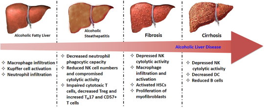
Ethanol is metabolised in the liver toacetaldehydeby alcohol dehydrogenase
which is in turn metabolised to acetate by aldehyde dehydrogenase.
As acetaldehyde is formed, NAD is metabolised to NADH as a cofactor in this reaction.
ThisNADH inhibits the actions of isocitrate dehydrogenase and alpha ketoglutarate and fatty acid oxidation, meaning it favours fatty acid synthesis.
Fat builds up in vacuoles of hepatocytes and mitochondria begin to dysfunction under oxidative stress, creating reactive oxygen species.
This fatty stage is known as steatosis and is stage one of alcoholic liver disease. It is reversible.
Stage Two
Inflammatory cytokines such as IL-6 and TNFa influx in, causing inflammation.
Water begins to accumulate inside hepatocytes and they begin to balloon. This causes significantcholestasis.
The inflammatory cytokines stimulate hepatocytes to produce collagen, which can begin to formfibrosis.
This collagen can be pericellular or around central veins, forming collagen bridges. This is a precursor for cirrhosis.
Stage Three
As the fibrosis progresses,hepatocytes begin to die and cirrhosis occurs.
This is end-stage alcoholic liver disease and can lead to liver failure and death.
Liver function tests
Instage 1; all LFTs will be raised. AST and ALT will be raised by approximately 5-10x the upper reference limit except albumin. They will both usually be below 300IU/L. AST will be raised above ALT; this is different to other hepatic conditions such as cholestasis, tumours, hepatitis, where ALT will be above AST.
In stage 2; ALP and bilirubin will increase significantly as cholestasis occurs and blocks bile duct. This will usually be above 300IU/L, which is different from filtrative diseases, where ALP is usually raised to 200-250IU/L. AST and ALT may increase further.
Instage 3; AST and ALT are between 10-100x the upper reference limit, suggesting significant cirrhosis and damage to cells. AST will remain above ALT. GGT will be raised significantly, which is common with alcoholic liver disease. Albumin will be decreased, suggestive of total liver failure. In other hepatic conditions, liver damage usually does not reach this stage and so albumin stays within the range.
Molar pregnancy is an abnormal form of pregnancy in which a non-viable fertilized egg implants in the uterus and will fail to come to term (will not develop into a child). Instead, the cells divide and replicate into a growing mass (mole) of non-foetal tissue.
Molar pregnancy is a gestational trophoblastic disease in which a non-viable egg grows into a mass (tumour) in the uterus that has swollen chorionic villi.
Can develop when a fertilized egg does not contain an original maternal nucleus.
Usually contains no foetal tissue.
Characterized by the presence of a hydatidiform mole (or hydatid mole).
Approximately 20% of women with a complete mole develop a trophoblastic malignancy (malignant disease // cancer)
Complete hydatidiform moles have a 2–4% risk of developing into choriocarcinoma in Western countries and 10–15% in Eastern countries, and have a 15% risk of becoming an invasive mole.
Molar pregnancies make up 1 in 1,000 pregnancies in the US and up to 1 in 100 pregnancies in parts of Asia.
Symptoms
Vaginal bleeding- molar tissue separates from the decidua, causing bleeding.
Uterus may become distended by large amounts of blood, and dark fluid may leak into the vagina.
Hyperemesis - severe nausea and vomiting due to very high levels of human chorionic gonadotropin (hCG).
Hyperthyroidism - thyroid gland is stimulated by the high levels of circulating hCG or by a thyroid stimulating substance (ie, thyrotropin) produced by the trophoblasts.
Partial mole
Partial moles do not generate the same clinical features as a complete mole. Patients instead present with signs and symptoms consistent with an incomplete or missed abortion, including vaginal bleeding and absence of foetal heartbeat.
Whole bone marrow cells (BMCs) and bone marrow mononuclear cells (BMMCs) are the most accessible and studied source of stem cells.
BMMCs are isolated from whole bone marrow, and contain a diverse cell population, including mesenchymal stem cells and hematopoietic progenitor cells.
Mesenchymal Stem Cells (MSCs)
MSCs can be isolated from a variety of tissues such as bone marrow, adipose, and umbilical cord; although it is not clear whether their properties are uniform (Selem, Hatzistergos and Hare, 2011).
MSCs are of particular note due to their immunoprivelegednature – a reduced expression of MHC class-I molecule, and lack of MHC class-II and co-stimulatory molecules, means they could potentially be used for allogeneic grafts (Zimmet et al., 2005). This means that they don’t produce an immune response and could be used in transplants - the body won’t reject them.
MSCs inhibit the activity of various immune cells, including T cells, B cells, natural killer cells, and dendritic cells via cell to cell contacts and soluble factors (Laflamme and Murry, 2005).
Foetal and Umbilical Cord Cells
Embryonic stem cells (ESCs), the prototypical stem cell, can develop into all cell types in the body. However, the practical application of human ESCs remains limited due to ethical problems, teratoma formation (cancer), and immune rejection. With rapidly expanding knowledge of molecular and genetic pathways for ESC differentiation, it may become possible to avoid contamination with undifferentiated ESCs, thereby inhibiting teratogenesis when transplanted into the body (Kucia et al., 2006).
Foetal-derived stem cells can also be isolated from the amniotic fluid, which include both pluripotent and committed stem cells.
Umbilical cord cells can be gathered at birth and stored, eg if for treatment later on if a defect is detected in utero.
Induced pluripotent stem cells (iPSCs)
Induced pluripotent stem cells are a more attractive alternative to ESCs, as they are autologous. This means cells can be taken from an individual, ‘reset’ back to their stem cell stage, and then administered to that same individual to avoid rejection. Pluripotency transcription factors are introduced to adult terminally differentiated somatic cells, such as dermal fibroblasts, in a novel strategy which ‘reprograms’ the cells back to an embryonic stem cell-like stage (Yu et al., 2007).
Despite slight epigenetic differences associated with reprogramming, iPSCs fully resemble ESCs in terms of differentiation capacity, morphology and gene expression profile; and have the ability to differentiate into other cells. Ethical and immune response dilemmas are bypassed by the autologous nature of iPSCs, however clinical application is not yet on the horizon due to their teratogenic potential and the oncogenes and virus vectors required for the current method of pluripotent induction (Yamanaka and Takahashi, 2006).
Skeletal myoblasts (SM)
Skeletal myoblasts (satellite cells) are derived from skeletal muscle and have the capacity to differentiate into muscle fibre, which makes them obvious candidates for treating conditions such as heart damage following infarction. However, clinical trials have been halted as SM have been observed to couple with resident cardiomyocytes, resulting in dysfunctional electrocardiology and arrhythmias, and have struggled to transdifferentiate into cardiomyocytes in vivo (Reinecke, Poppa and Murry, 2002).
Stem cells are cells in the body that don’t yet have any role (undifferentiated or partially differentiated), and can change to become almost any cell type.
Canproliferate (divide to make more cells) indefinitelyto make more of the same stem cells.
They are the earliest type of cell in a cell lineage (if a cell was an adult human, stem cells would be the foetus)
found in both embryonic and adult organisms, but they have slightly different properties in each
Can also be made in a lab by reprogramming other cells - resetting them back to stem cell stage.
Inembryonic development (a baby forming in the womb), pluripotent stem cells develop at the blastocyst stage (3-4 days) and differentiate into all the cells of the human body as the foetus develops.
Stem cells do exist in the adult body, however they are not pluripotent - they are unipotent or multipotent - this means they can only differentiate into one or a few cell types respectively.
Adult stem cells
Adult stem cells are found in a few select locations in the body, known as niches, such as:
the brain
bone marrow
blood and blood vessels
skeletal muscles
liver
gonads
They exist to replenish rapidly lost cell types and include hematopoieticstem cells, which replenish blood and immune cells,basal cells, which maintain the skin epithelium, and mesenchymal stem cells, which maintain bone, cartilage, muscle and fat cells. Adult stem cells are a small minorityof cells.
Malignant lesions of the ovaries include primary lesions arising from normal structures within the ovary and secondary lesions from cancers arising elsewhere in the body.
Signs and symptoms
Bloating; abdominal distention or mass
Pressure effects on the bladder and rectum
Constipation, indigestion, reflux
Vaginal bleeding
Shortness of breath, tiredness, weight loss
Diagnosis
Physical findings are uncommon in early stages. Advanced disease may present with ovarian or pelvic mass, ascites, pleural effusion, or abdominal mass or bowel obstruction.
Pathophysiology
Typicallyspreads to the peritoneal surfaces and omentum.
Occurs via local extension, lymphatic invasion, intraperitoneal implantation, hematogenous dissemination, or transdiaphragmatic passage.
Malignant cells can implant anywhere in the peritoneal cavity but are more likely to implant in sites of stasis along the peritoneal fluid circulation.
Epithelial tumours make up 90% of ovarian tumours. Other histologies include:
Sex-cord stromal tumors
Germ cell tumors
Primary peritoneal carcinoma
Metastatic tumors of the ovary
Epithelial ovarian cancer
Arises from epithelium covering the fimbria of the fallopian tubes, or the ovaries, both of which are derived from the coelomic epithelium.
Found primarily as cystic lesions with solid components.
Surface may be smooth or covered in papillary projections.
Cysts contain fluid from yellow to brown and haemorrhagic.
Four main histologic subtypes:
Serous (from fallopian tube)
Endometrioid (endometrium)
Mucinous (cervix)
Clear cell (mesonephros)
Tumours of low malignant potential
Tumours of low malignant potential (LMP) are a variety of much less aggressive epithelial ovarian cancer, with good prognosis.
LMP tumors can cause a range of symptoms similar to epithelial ovarian cancer, including increasing abdominal girth, an abdominal mass, abdominal pain, abnormal uterine bleeding, urinary symptoms, and gastrointestinal symptoms. They may be asymptomatic and found on routine physical examination or ultrasound scan.
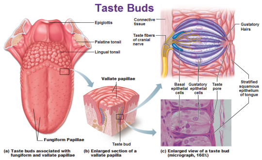
The tongue is covered with many little bumps called papillae.Taste buds are found in the walls of papillae and the grooves surrounding them. Each taste bud contains anywhere from 50 to 150 taste receptor cells.
Microvilliextend from taste receptor cells
and protrude through an opening (taste pore) into the mouth.
These microvilli come in contact with substances in the mouth that can be tasted, also known as tastants.
Tastants interact with taste receptor cells through a number of different mechanisms to depolarize the cells.
When taste cells are depolarized, they release neurotransmitters that stimulate sensory neurons that travel in cranial nerves VII, IX, and X.
These neurons terminate on neurons in the nucleus of the solitary tract in the medulla then continue on to the thalamus.
Taste information is sent to the gustatory cortex, ( ocated on the border between the anterior insula and the frontal operculum).
This information encodes for basic tastes, such as sweet, salty, sour, bitter, and savory or umami.
However, the actual flavour of a food—which is what we typically define as taste—is created by a combination of taste and olfactory (smell) information.
Sweetness
Produced by the presence of sugars, some proteins, and other substances.
Detected by G protein-coupled receptors T1R2+3 (heterodimer) and T1R3 (homodimer).
Saltiness
Saltiness is a taste produced best by the presence of cations (such as Na+, K+or Li+)
Directly detected by cation influx into glial like cells via leak channels causing depolarisation of the cell.
Sourness
Sourness is acidity and is also sensed using ion channels.
Undissociated acid diffuses across the plasma membrane of a presynaptic cell, where it dissociatesin accordance with Le Chatelier’s principle.
Theprotons that are released then block potassium channels, which depolarise the cell and cause calcium influx.
Bitterness
Current research suggests TAS2Rs (taste receptors, type 2, also known as T2Rs) such as TAS2R38 are responsible tasting bitter substances.
Savouriness
The amino acid glutamic acid is responsible for savouriness, but some nucleotides (inosinic acid and guanylic acid) can act as complements.
Glutamic acid binds to a variant of the G protein-coupled receptor, producing a savoury taste
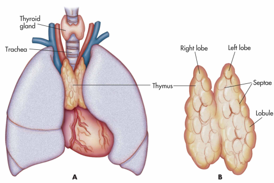
Thethymus is a specialised primary lymphoid organ of the immune system.
At its largest and most active during the neonatal and pre-adolescent periods.
Decreases in size and activity through teenage years
Thymus tissue is gradually replaced by adipose tissue(fat).
Residual T lymphopoiesis continues throughout adult life.
The thymus is composed of two identical lobes and is located in the anterior superior mediastinum, in front of the heart and behind the sternum. Each lobe of the thymus can be divided into a central medulla and a peripheral cortex which is surrounded by an outer capsule.
Function
Facilitates the maturation of T cells - which provide cell-mediated immunity.
T cells begin as hematopoietic precursors from the bone-marrow, and migrate to the thymus, where they are referred to as thymocytes.
In the thymus they undergo a process to ensure the cells react against antigens (“positive selection”), but that they do not react against antigens found on body tissue(“negative selection”).
Once mature, T cells emigrate from the thymus to provide vital functions in the immune system.
Each T cell has a distinct T cell receptor, suited to a specific substance, called an antigen.
Most T cell receptors bind to the major histocompatibility complex on cells of the body.
Positive selection
T cells have distinct T cell receptors. These are formed by process recombination gene rearrangement which is error-prone, and some thymocytes fail to make functional T-cell receptors, whereas other thymocytes make T-cell receptors that are autoreactive. The survival and nature of the T cell then depends on its interaction with surrounding thymic epithelial cells.
T cell receptor interacts with the MHC molecules on the surface of epithelial cells.
A T cell with a receptor that doesn’t react, or reacts weakly will die by apoptosis.
A T cell that does react will survive and proliferate.
A mature T cell expresses only CD4 or CD8, but not both.
Negative selection
T cells that attack the body’s own proteins are eliminated in the thymus. Epithelial cells in the medulla and dendritic cells in the thymus express major proteins from elsewhere in the body. Some CD4 positive T cells exposed to self antigens persist as T regulatory cells.
Pathology
Immunodeficiency - As the thymus is the organ of T-cell development, any congenital defect in thymic genesis or a defect in thymocyte development can lead to a profound T cell deficiency in primary immunodeficiency disease.
Autoimmune disease - Genetic disorders, such as Myasthenia gravis: caused by antibodies that block acetylcholine receptors.
Thymomas - Originate in thymic epithelial cells most often in adults older than 40. Generally detected when they cause symptoms, such as a neck mass or affecting nearby structures such as the superior vena cava. Can be benign; benign but by virtue of expansion, invading beyond the capsule of the thymus (“invasive thyoma”), or malignant (a carcinoma).
Lymphomas - Tumours originating from T cells of the thymus form a subset of acute lymphoblastic leukaemia (ALL)
Thymic cysts - The thymus may contain cysts, usually less than 4 cm in diameter. Thymic cysts are usually detected incidentally and do not generally cause symptoms.
Y'all ever just suddenly have the overwhelming urge to swim??? Like not actively but you just wanna,,, be in the water and have some Peace
Yes it’s called the mammalian diving response and it’s also why doing face masks and taking a shower is soothing. Our amphibian ancestors used this mechanism to slow down the heartbeat and lower body temperature so as not to waste calories while swimming (which is very calorie intensive). It makes you feel safe because predators are less likely to get you in water than on land. The fish brain is alive and well in all of us.
It’s literally activated by putting water on the face.
Very proud of our Gallery of Humankind, our showroom of human life: you can explore our 7 million years of human past and - as you see here - the human body. Studio Louter made these beautiful projections.
This zone openly and honestly explores different life stages, from the embryo to adulthood: fertilisation, pregnancy, birth and the first weeks of life, a child’s rapid growth, the changes that take place during adolescence (to the brain and future reproductive functions) and old age.
Biological sex is not and has never been a binary. The complexity of the natural world cannot be contained in neat little societal boxes. Stop using science to justify your bigotry.
The complexity of the natural world cannot be contained in neat little societal boxes. Stop using science to justify your bigotry.
Can you even imagine being the poor alien sod responsible for auditing an earthling spaceship’s spending allowance? Like:
“I see, and why do you require many tubes of white plant flavoured paste?”
“Oh well, if we don’t rub that on our teeth twice daily the bacteria living in my mouth will begin to devour me teeth.”
“…Noted.”
“I have also noticed several large shipments of specific medications, and a variety of individually packaged absorbent material - however injury records do not show sufficient numbers to justify these recurrent deliveries.”
“Ah, yeah, it’s not really an injury per say. As part of our natural reproductive cycle approximately half the population will shed the lining of one of their internal organs and expel it.”
“…that is the most horrifying thing that I have ever heard.”
“Yeah.”
“Does such a process not hurt?”
“That’l be what the medication’s for. Pain killers for the cramps, birth control to stop the process.”
“…and your reasoning behind the fully functional, high-tech entertainment system?”
“Okay, that we could probably do without. But in our defence that was actually insisted on as a standard feature of all fleet-ships expected to encounter Terrans. Admiral Plo’Kaght insisted on it. Something about bored humans and a an illegal betting ring featuring a cleaning robot with a knife strapped to it going up against a human with a mop?”