#histopathology

Ghostface Killer is back!
Only this time he’s hiding in a rectum!
Scaring while you’re learning!
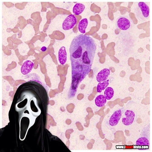
A rectal scrape is performed (more often in veterinary med) to obtain a sample of the epithelial cells lining the rectum. These are observed in the microscope for abnormal changes or for microbes and their eggs/larvae that might be present in the digestive tract.
This particular scrape contains an epithelial cell that appears to be bi-nucleate and is surrounded by red blood cells.

Gingerbread Placenta
Run, run, run as fast as you can
You’ can’t catch me, I’m the chorionic villus gingerbread man!
The image shows a section through one of the many thousands of chorionic villi in the placenta that are responsible for the exchange of gas and nutrients with the maternal blood.
The mostly white space are the maternal blood lakes which are normally filled with mom’s blood while the small vessels (like gingerbread’s eyes and mouth) within the villus are branches of the umbilical vessels that shuttle blood back and forth to and from the growing baby.
The very thin cells lining the villus (gingerbread’s skin) are syncytiotrophoblast cells which gas must diffuse across in order to move from mom to baby and vice versa.
Histology by @BiopsyMD via Twitter

Mother Earth
A biopsy of the mammary gland obtained during pregnancy.
Let’s celebrate our planet and home!
Happy Earth Day everyone!
i♡histo
Post link