#med school


HAI friends it me again, posting what I love posting, a handwriting closeup :D this is from our very first surgical lecture so it made me really happy to make them, I’m starting to feel like a real vet..
I’m working on yet another youtube video (omg they are so much FUN to make let me tell you) but if you want to check out my last one where I talk about vet school, link is: https://www.youtube.com/watch?v=Z7C7dWX_hvs
as usual, I love hearing from you guys so feel free to send me asks or messages or coomments or whatever it is the kids do these days about how you’re doing and how your weeks are going <3 also I’m headed to a cat cafe later might spam everyone with cat pics sorry not sorry kbyeloveyou
♫ Seventeen, No Rome
Post link

“After the kids left the house, I decided to go to medical school and become a doctor. I was 48 at the time, and had been out of school for a long time. When I asked him if he thought it was a good idea, he said: ‘Sure. It’s your turn.’”
Post link

Late night quesmed :)

✨ Happy Star Wars Day ✨
May the Fourth be with you!
i♡histo
Star Wars histology from top left, clockwise:
1. The Graafian follicle Death Star
In a galaxy far, far away an intergalactic superweapon is halted in metaphase II of meiosis amid a surge in Luteinizing Hormone.
2. Jabba the Corpus Albicans
“makingsa lee ka bok pateesa… beeska chata wnow kong bantha poodoo”
(translation) “you may have been a good friend..but now you are bantha fodder”
The corpus albicans is a structure in the ovary that is formed when the corpus luteum regresses.
3. Tusken Raider in the Liver
Despite what you see here, Tusken Raiders are not native to the human liver. If you think that, then you are making a wookie mistake.
The image is actually a portal triad and demonstrates the major structures that enter and leave the liver: hepatic artery, hepatic portal vein and bile duct.
4. The Empire Strikes Back (at the Liver)
Liver histology is definitely where it’s At-At!
A region of connective tissue among the hepatocytes in the liver.
—
Images by @ihearthisto,@drjohnrajala,@zenonichand@hopkins_gi_pathrespectively
Post link

Esoph-egg-us
✅Non-keratinized stratified squamous epithelium
✅Circumscribing muscularis mucosae
✅Submucosal glands
✅Dual layered muscularis externa
It’s just not Spring until you have painted your very own Esoph-egg-us
So get cracking and I don’t want to hear any eggscuses
#histology #science #pathology #pathologists #anatomy #autopsy #eggs #easter #spring #biology #esophagus #digestive #premed #meded #nurse #nursing #medschool #medstudent #medicine #education #vetscience #vetschool #dentistry #histotechnology #histologica #histotech #histo #pathArt #sciArt #ihearthisto

Grumpy Cord
A transverse slice through a spinal cord that looks like Grumpy Cat.
Being the center of the nervous system, transmitting neural signals between the brain and the entire body and controlling independent neural reflexes…is just so awful .
The pale central face is the ‘grey matter’ and is home to the many neuron cell bodies that run through the spinal cord. The surrounding darker region is composed of the axons of these neurons as the exit, enter, ascend and descend through the spinal cord on their way to innervate muscles, or returning information about pain back from the skin, or relaying information about body position back to the brain.
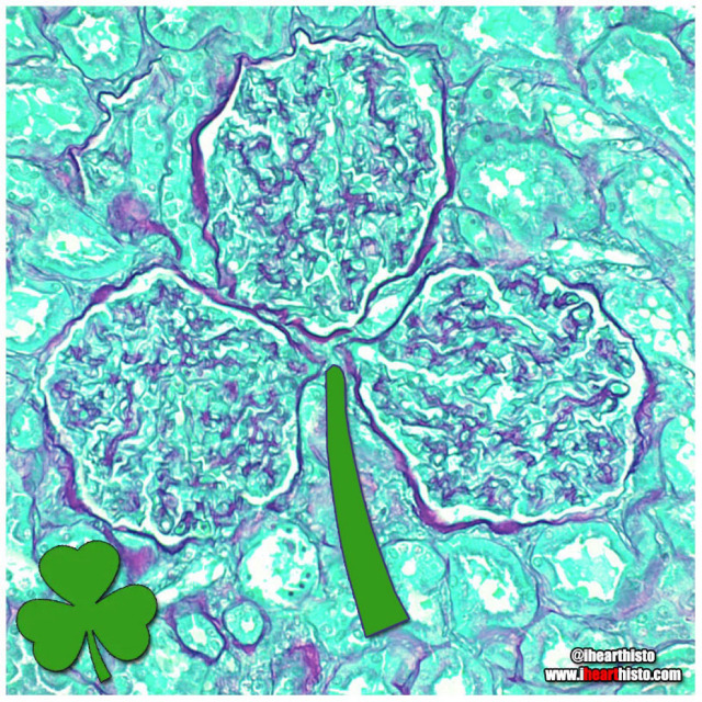
☘️Shamrock Gloms ☘️
For each petal on the shamrock,
This brings a wish your way.
Good health. Good luck. Happiness.
For today and every day.
Happy St. Patrick’s Day!
Three renal corpuscles (glomerulus + their surrounding Bowman’s capsules) floating in a sea of distal and proximal convoluted tubules within the cortex of the kidney.
These three small structures are knotted balls of capillaries (glomeruli) surrounded by a specialized epithelium (Bowman’s capsule) that is composed of cells called podocytes. These cells have tiny interlocking legs that form a small slit between them.
This structural organization is responsible for filtering your blood to produce a fluid that then travels within tubes continuous with the Bowman’s capsule called nephrons. In these nephrons the tubular fluid is modified by reabsorbing and secreting ions and conserving water to produce urine for excretion.

⚡Lord Voldermis⚡
A biopsy of a region of skin-that-shall-not-be-named (dermis/hypodermis junction shhh), complete with nerves, vessels, sweat glands and hair follicles.
by @nejiby

You can find LOVE in the strangest of places (2022 edition)
By row starting top left:
1. in a skin cylindroma
2. in an hepatic ductule
3. in a pancreas
4. in a warty penile growth
5. in a mucus-y colon
6. in a region of hypodermis
7. in a secondary oocyte
8. in a chondrosarcoma
9. in a small artery
Happy Valentine’s Day
Tag a friend with the histo heart you want to share with them and spread the love!
Images by:
@ihearthisto [1-4, 5-9]
@donna.horncastle [5]

Kitty Liver
Check meowt!
I’m a purrfect hepatic vein surrounded by hepatic lobules!
This is a hematoxylin and eosin stained slice through a liver that was injected with carbon/ink prior to fixation.
The liver is compose of numerous roughly hexagonal shaped lobules. Each lobule has a venule at its center (the central vein; top right) and a series of hepatic triads at its periphery. A triad is a collection of three structures - in this case branches of the hepatic artery, hepatic portal vein and hepatic duct.
Blood in the hepatic artery (oxygenated) and hepatic portal vein (rich in nutrients absorbed from the intestines) travels in sinusoids (the many narrow white spaces in this image) towards the central vein of each lobule. On its way through the sinusoids the blood is processed by the the many hepatocytes that line this region (the pink cells).
Within the sinusoids reside many liver macrophages (Kupffer cells) that phagocytose debris traveling through sinusoids. Normally these cells are invisible in standard H&E preparations but recall that this tissue was injected with ink! The ink was phagocytosed by the macrophages filling their cytoplasm with carbon so that they are now visible as black cells in the sinusoids.
Once the blood enters the central vein of each hepatic lobule they drain into the hepatic veins (kitty!) until the blood reaches the inferior venal cava.
In this way all blood, rich in raw nutrients and toxins absorbed from the GI tract is processed by the liver before it enters the systemic circulation!
Pawsitively unbeliverable!
Post link
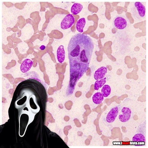
Ghostface Killer is back!
Only this time he’s hiding in a rectum!
Scaring while you’re learning!
A rectal scrape is performed (more often in veterinary med) to obtain a sample of the epithelial cells lining the rectum. These are observed in the microscope for abnormal changes or for microbes and their eggs/larvae that might be present in the digestive tract.
This particular scrape contains an epithelial cell that appears to be bi-nucleate and is surrounded by red blood cells.

Cookie Monster Ovary
Today’s ihearthisto is brought to you by the letter O for Oocyte!
This is image shows a high magnification view of a slice through an ovary.
You are looking at four tiny primordial follicles (the cookies above Cookie Monster’s head) located in the outer cortex of the ovary. Each of these follicles contains a single dormant, immature egg (a primary oocyte) that is halted in the the first phase of meiosis (prophase I). Eventually these follicles will be recruited to enter the maturation cycle that, against all the odds, could see them develop into an embryo.
Cookie monster himself is a larger multilaminar primary follicle (the cookie in his mouth is the nucleus of the oocyte within the follicle). This type of follicle has already been recruited and it’s follicular cells are dividing and differentiating. The oocyte within it though is still halted in prophase I of meiosis.
An oocyte will only complete meiosis if it is ovulated and fertilized by a spermatozoon (a sperm cell).
Post link
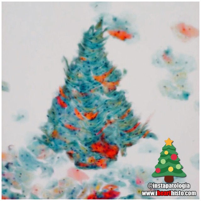
❄️Pappy Holidays❄️
There’s nothing quite like a Pap smear Christmas tree to rock around this happy holiday season!
by the awesome @instapatologia [Insta]

Have a Howelly-Jolly Christmas
A festive finding in the blood of an asplenic patient
A Howell–Jolly body is a cytopathological finding whereby small remnants of nuclear DNA are present in normally anuclear circulating erythrocytes.
During development in the bone marrow, late orthochromatophilic erythroblast normally expel their nuclei. However, in some cases, a small portion of DNA remains (the purple dots in the erythrocytes wearing the Santa hats).
Under normal circumstances if these irregular erythrocytes make it into the blood, they are removed from circulation by the spleen.
As a result, the presence of erythrocytes with Howell-Jolly bodies in peripheral blood smears like this usually signifies a damaged or absent spleen - because a healthy spleen would normally filter this type of red blood cell.
by exlibrisadpugno via reddit

Gingerbread Placenta
Run, run, run as fast as you can
You’ can’t catch me, I’m the chorionic villus gingerbread man!
The image shows a section through one of the many thousands of chorionic villi in the placenta that are responsible for the exchange of gas and nutrients with the maternal blood.
The mostly white space are the maternal blood lakes which are normally filled with mom’s blood while the small vessels (like gingerbread’s eyes and mouth) within the villus are branches of the umbilical vessels that shuttle blood back and forth to and from the growing baby.
The very thin cells lining the villus (gingerbread’s skin) are syncytiotrophoblast cells which gas must diffuse across in order to move from mom to baby and vice versa.
Histology by @BiopsyMD via Twitter

Rudolph the Red-Nosed Reindeer
Had a syncytiotrophoblast nose
And if you ever saw it
You would even say: “it forms the placental surface across which gases, nutrients & metabolites pass from the maternal circulation to enter the fetal circulation & vice versa”
This image shows many slices through the placental villi, fingerlike projections of the fetal-derived component of the human placenta. One of these villi looks like he’s been prohibited from participating in a variety of reindeer activities.
Rudolph’s core is composed of mesenchyme, an embryonic tissue that has the capacity to form tiny vessels (the tiny white holes) that are branches and tributaries of the umbilical arteries and vein that run in the umbilical cord and hook up with baby’s internal vascular plumbing.
The cell layer forming Rudolph’s skin (and his nose!) is called the syncytiotrophoblast layer. In the early placenta there are actually two layers (the outer syncytiotrophoblast and the inner cytotrophoblast). As the placenta matures the cytotrophoblasts thins and disappears leaving only the syncytiotrophoblast as the thin barrier between moms blood and those tiny fetal capillaries inside each villus.
But where is mom’s blood I hear you ask? Well, you can’t see it because it drained away once the placenta was removed and was sliced up. But… what you can see are the large ‘maternal blood lakes’ where her blood used to be (the large white spaces).
Now imagine all those fetal villi, and Rudolph, floating in those blood lakes and you can start to appreciate how efficient this arrangement is at allowing the exchange of gases and nutrients between mom and baby.

Prostate Snail
A secretory gastropod with a corpus amylaceum shell!
i❤️histo
This image is a close up view of a slice through the prostate gland!
The glandular portion of the prostate is composed of secretory acini that release prostatic fluid into a duct system within the gland. Prostatic fluid is a major component of semen and is rich in protein and sugar that keeps spermatozoa nourished as they travel through the reproductive tract.
The snail’s head and body are composed of the secretory epithelial cells!
The giant shell of the snail is a structure known as a prostatic concretion (corpus amylaceum or starchy body). This is a substance thought to be composed of thickened prostatic secretions and shed cells that is found in the acini and ducts of the prostate gland - it is of unknown significance.
However, these structures do increase in number with age and are a useful identifying feature of prostate in both non-pathological and pathological prostate specimens.
The space between the secretory acini is filled with a mixture of fibrous connective tissue and smooth muscle.
The smooth muscle is important because it contracts during ejaculation to push the prostatic fluid out of the gland and into the prostatic urethra where it mixes with spermatozoa arriving from the testis.
The connective tissue component is important clinically because as people with prostate glands grow older this fibrous tissue can undergo hyperplasia (excessive growth). This growth can constrict the urethra which passes directly through the prostate gland. The urethra, in addition to conveying semen during ejaculation, also carries urine during micturition/urination. This explains some of the symptoms associated with the common disorder, benign prostatic hyperplasia when urination becomes problematic for the elderly.
Post link
Students:
Histology:
- - - -
Those basic tissues are tough. But you have got this.

VERYSmooth Muscle
i♡histo

The Cytology Zoo
You can come too, too, too!
An exhibition of smears featuring some familiar looking squamous epithelial cell critters.
Can you recognize them all?
Cytology is the study of individual cells that have been isolated from a specimen. The technique is used mainly to study or screen for cancer but can be useful in the diagnosis of other diseases too, like those involving infectious organisms.
Most of the cells seen in a cytological smear or specimen are obtained in one of three ways:
- Scraping/Brushing the surface of a tissue (this is how cells are obtained from the uterine cervix during a pap smear)
- Collecting a fluid, this could be urine, mucus, blood or semen
- Fine-needle aspirations, this is when cells are removed by sucking them through a fine diameter needle. Fluid from the abdominal, pleural or pericardial cavities are obtained in this way and also cerebrospinal fluid during a lumbar puncture (or spinal tap).
The cells seen here are all squamous epithelial cells that are obtained from the cervix by scraping/brushing.
Credits:
@oreoimc (1-4) & (7-9)
@isac.p (5)
@sza_jhycto (6)
#histology #science #pathology #pathologists #anatomy #autopsy #zoo #animals #cytology #dentalschool #iquizhisto #premed #biology #medicaleducation #meded #nurse #nursing #medschool #medstudent #medicine #medlab #vetscience #vetschool #vetstudent #histologia #histotech #histo #pathArt #ihearthisto