#histotech
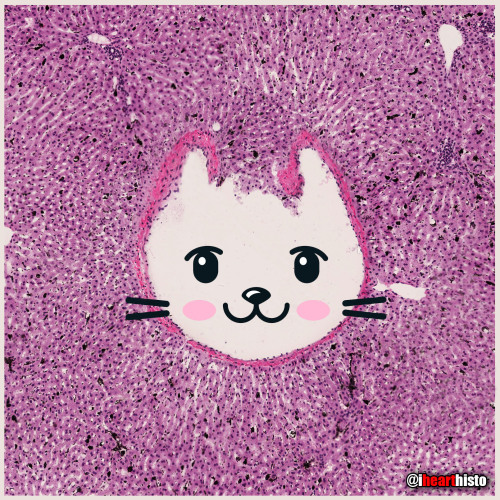
Kitty Liver
Check meowt!
I’m a purrfect hepatic vein surrounded by hepatic lobules!
This is a hematoxylin and eosin stained slice through a liver that was injected with carbon/ink prior to fixation.
The liver is compose of numerous roughly hexagonal shaped lobules. Each lobule has a venule at its center (the central vein; top right) and a series of hepatic triads at its periphery. A triad is a collection of three structures - in this case branches of the hepatic artery, hepatic portal vein and hepatic duct.
Blood in the hepatic artery (oxygenated) and hepatic portal vein (rich in nutrients absorbed from the intestines) travels in sinusoids (the many narrow white spaces in this image) towards the central vein of each lobule. On its way through the sinusoids the blood is processed by the the many hepatocytes that line this region (the pink cells).
Within the sinusoids reside many liver macrophages (Kupffer cells) that phagocytose debris traveling through sinusoids. Normally these cells are invisible in standard H&E preparations but recall that this tissue was injected with ink! The ink was phagocytosed by the macrophages filling their cytoplasm with carbon so that they are now visible as black cells in the sinusoids.
Once the blood enters the central vein of each hepatic lobule they drain into the hepatic veins (kitty!) until the blood reaches the inferior venal cava.
In this way all blood, rich in raw nutrients and toxins absorbed from the GI tract is processed by the liver before it enters the systemic circulation!
Pawsitively unbeliverable!
Post link
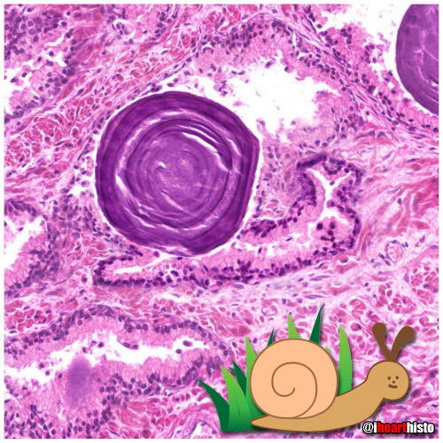
Prostate Snail
A secretory gastropod with a corpus amylaceum shell!
i❤️histo
This image is a close up view of a slice through the prostate gland!
The glandular portion of the prostate is composed of secretory acini that release prostatic fluid into a duct system within the gland. Prostatic fluid is a major component of semen and is rich in protein and sugar that keeps spermatozoa nourished as they travel through the reproductive tract.
The snail’s head and body are composed of the secretory epithelial cells!
The giant shell of the snail is a structure known as a prostatic concretion (corpus amylaceum or starchy body). This is a substance thought to be composed of thickened prostatic secretions and shed cells that is found in the acini and ducts of the prostate gland - it is of unknown significance.
However, these structures do increase in number with age and are a useful identifying feature of prostate in both non-pathological and pathological prostate specimens.
The space between the secretory acini is filled with a mixture of fibrous connective tissue and smooth muscle.
The smooth muscle is important because it contracts during ejaculation to push the prostatic fluid out of the gland and into the prostatic urethra where it mixes with spermatozoa arriving from the testis.
The connective tissue component is important clinically because as people with prostate glands grow older this fibrous tissue can undergo hyperplasia (excessive growth). This growth can constrict the urethra which passes directly through the prostate gland. The urethra, in addition to conveying semen during ejaculation, also carries urine during micturition/urination. This explains some of the symptoms associated with the common disorder, benign prostatic hyperplasia when urination becomes problematic for the elderly.
Post link
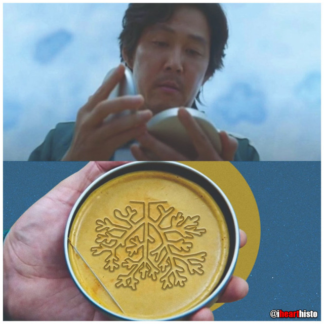
Students:
Histology:
- - - -
Those basic tissues are tough. But you have got this.

VERYSmooth Muscle
i♡histo

Bender-scope
The microscopes of the future are all surly and a little bit hungover.
Current mood: “Bite my shiny metal a$$“
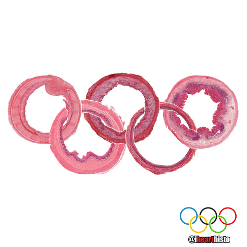
The Organ Rings [Answers]
Original post without answers:
https://www.ihearthisto.com/post/657148528765911040/the-organ-rings-to-commemorate-the-olympic
Trachea: C-shaped cartilage, a pseudostratified epi with cilia & goblet cells
Esophagus: Non-keratinized stratified squamous epi and muscularis mucosa
⚫ Artery (distributing): Prominent internal elastic lamina & thick smooth muscle tunica media
Bladder or Ureter: Transitional epithelium
Appendix: Crypts of Lieberkuhn (no villi) with surrounding submucosal lymph nodules
Thanks for playing!
Enjoy the games!
Post link
The Organ Rings
To commemorate the Olympic Games here are some slices through a few of the more tubular organs in your body!
Do you know what they are? Comment below!
Winner gets the gold medal!
[I’ll post the answers soon!]
Post link

❤️ Thanks Dad!
These are real spermatozoa.
Incidentally, based on head morphology, they are probably from a bull and not a human.
They were observed and photographed using phase contrast microscopy, a technique commonly used in semen analysis.
Note that the spermatozoa were realigned using photo editing software - spermatazoa are pretty cool cells but they do need a little help with their spelling sometimes.
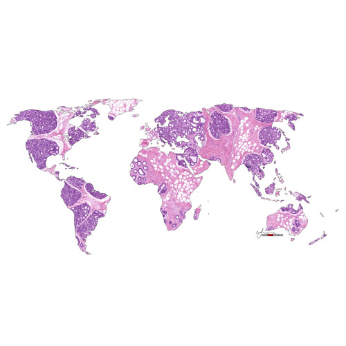
Mother Earth
A biopsy of the mammary gland obtained during pregnancy.
Let’s celebrate our planet and home!
Happy Earth Day everyone!
i♡histo
Post link
The Ro-lung Stones
The Stones’ ‘Hot Lips’ logo formed from a blood vessel filled with erythrocytes within a congested zone around a focal pneumonia of the lung.
During the first stages of focal/lobar pneumonia macrophages (the larger cells that are visible in the white alveolar space) respond to phagocytose (eat) any pathogens in the lung.
Additionally, the small blood vessels within the lung tissue begin to engorge (you can see all the erythrocytes in the dilated vessels not only in the large 'hot lips’ vessel but the smaller vessels in the interalveolar septa between adjacent alveoli). This is the congestion phase.
With this dilation of vessels come more white blood cells, you can see numerous neutrophils (they are the small cells that look like they have multiple lobes to their purple nuclei) have migrated out of the vessels (a process called extravasation) into the surround tissue and alveolar spaces. These cells are signs of an additional immune response and they help the macrophages destroy pathogens in the airway.
Image is by @beautiful_pathologist - check out her Instagram for more histology.
Post link

Happy Vagina Scalp Epididymis Liver!!
Here’s to a healthier, happier New Year for everyone.
Image shows:
2 - Blood vessel in vaginal mucosa
0 - Hair follicle in scalp
2 - Epididymis
1 - Hepatic portal vein branch in liver
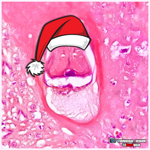
Santa Larvae
Hurry down my cecum tonight!
A nematode larva (Contracaecum) in the digestive tract of a seal. The eggs of this parasitic nematode use fish as an intermediate host before infecting piscivorous mammals, including humans who may forget to clean their holiday salmon!
This festive image was captured by veterinary pathologist @velphegor_vetpath via Instagram.
Happy holidays everyone!
Post link
Blobfish-face
A coronal section through a developing jaw (when flipped upside down) looks like poor old blobfish!
The blobfish eyes are formed from Meckel’s cartilage. A component of the first pharyngeal arch that runs the length of the developing mandible. It degenerates as the fetus develops leaving only two small components on each side of the head. These ossify (become bone) to form the incus & malleus (ear ossicles) of the middle ear.
The blobfish nose is the developing tongue. It is composed of developing skeletal muscle fibers. Skeletal muscle forms from myoblasts that line up & fuse to form long myotubes. These will then synthesize actin/myosin which will allow them to contract & form the intrinsic skeletal muscle of the tongue.
The blobfish chin is formed by the developing maxilla. Two regions of tissue (the palatine shelves) grow together & fuse in the midline to form the posterior hard palate. You can see the midline suture forming & feel it in your own mouth by running your tongue along the roof of your mouth. Failure of these to fuse results in a variety of cleft lip and palate combinations.
The blobfish head is formed from a developing mandible. You can see small islands of bone forming within the mesenchymal tissue of the head. This type of bone development is called intramembranous ossification.
The blobfish is native to coastal waters off mainland Australia and Tasmania where it lives way deep down in the darkest depths of the ocean. Its gelatinous body is ideal for withstanding the pressure down there but when brought to the surface it looks like a sad melted pink crayon.
#histology #science #pathology #pathologists #anatomy #autopsy #blobfish #embryology #premed #biology #dentalschool #dentalstudent #dentistry #medicaleducation #meded #nurse #nursing #medschool #medstudent #medicine #medlab #vetscience #vetschool #vetstudent #histologia #histotech #histo #pathArt #ihearthisto
Post link
Caaaaarrrggghhh-diovascular Histology
Terrifying a medical student near you this Halloween!
The zombie’s lifeless eyes are arterioles.
His blood-filled mouth is a small vein.
All wrapped up in a connective tissue face.
Happy Halloween Histo fans!
Post link
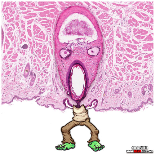
Zomb-hair
A very close-up scene from the hit TV series ‘The Walking Dreads’
This is actually a vibrissae hair follicle from the face of a (a whisker).
Vibrissae are sensory hairs that are different from regular body hairs in the fact that they are surrounded by a blood filled sinus (z’s brain) and are associated with sensory neurons that have a distinct and representative pattern in the somatosensory cortex of the mouse brain. The fact that they are well mapped in the brain illustrates their importance in everyday behavior and survival - they are involved in things like detecting, orienting and sensing length of surfaces/objects and tracking (e.g. finding gaps in a maze and answering the question 'can my head fit through here?’).
Note also the sebaceous glands (z’s eyes) which secrete a lipid rich secretion called sebum for maintaining hair and epidermis keratin (keratin is the wispy stuff lining the skin at the bottom of the image and forming z’s lil arms).
The hair in this follicle is absent as you can see z’s wailing mouth is completely empty (where you might expect to see the shaft of a hair).
The closest things us humans have to vibrissae are the thick long hairs in our nostrils which are fairly sensitive to tickles (and make your eyes water if plucked) but more importantly provide a first line of filtration to prevent big particles from being inhaled into your nasal cavity.
#histology #science #anatomy #pathology #autopsy #pathologists #dermatology #halloween #hair #skin #zombie #dentalstudent #dentalschool #histotech #medlab #premed #meded #nurse #nursing #medschool #medstudent #medicine #vetscience #vetschool #histotechnology #histologia #histo #pathArt #vetstudent #ihearthisto
Post link
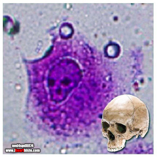
The Creepiest Fibroblast You Will Ever See
With a nucleus straight from the fiery depths of hell*
*Note: Fibroblasts do not come from hell. They are actually derived from embryonic mesoderm and the creepiest thing they do is secrete the collagen and ground substance of connective tissue. Not very creepy at all really when you think about it.
Histology is from the microscope of the ‘fiancee of ned4spd8834’
Post link

![The Organ Rings [Answers] Original post without answers: https://www.ihearthisto.com/post/657148528 The Organ Rings [Answers] Original post without answers: https://www.ihearthisto.com/post/657148528](https://64.media.tumblr.com/484b98cf82c61d709722bca340d7cc7e/23e52e0dade3875d-bb/s500x750/6f5285a59611a5ae98534a85ae3cf72dfaedab18.jpg)
