#vet science


BIOCHEMISTRY BLOOD TESTING
Biochemistry blood test measures the levels of chemical substances carried in the blood. This type of test allows us to evaluate the how well the liver and kidneys are working and how much fat and sugar is circulating in the bloodstream.
Blood Glucose
When carbohydrates are eaten they are broken down and stored in the Liver as Glycogen until the animal needs energy where it is then converted to glucose and transported around the body. We use blood glucose as a monitor of metabolism and physiology.
Normal BG (Canine) - 5.6 to 13.9mmol/L
Normal BG (Feline) - 5.6 to 16.7mmol/L
INCREASED BG - Diabetes Mellitus is indicated however it is recommended that the urine is also checked for Glucose as if present this means the kidneys have reached their threshold and Diabetes is very likely. Note that cats can have stress induced hyperglycemia and so a diagnosis of diabetes should not be made on a single BG reading.
DECREASED BG - Patients that are sick and deliberated often have hypoglycemia. but puppies who have been starved for procedures can also suffer from a low BG. In addition to this hunting breeds that have been working hard for a prolonged time can also suffer from a low BG.
Bun Urea Nitrogen (BUN)
BUN is the by product produced when Proteins are broken down and used within the body. This by-product is excreted by the Kidneys in the urine.
INCREASED BUN - This would mean the kidneys are not working sufficiently and could be an indicator of kidney disease or kidney obstruction that is preventing urine reaching the bladder and therefore build up in the Kidney. Heart disease causing poor circulation to the kidneys could also be a cause of increased BUN.
DECREASED BUN - As the liver breaks down Protein a lower level of BUN could indicate that the liver is not working as well as it should and isn’t breaking down protein as well as it should.
Creatinine (CREA)
Creatinine is solely filtered out of the blood by the kidneys.
INCREASED CREA - Impaired Kidney function
Calcium (CAL)
Calcium is a mineral that is found at a consistent level within the blood. It’s needed for muscle and nerve function and without it death can occur.
INCREASED CAL - Some types of cancers and medications can cause an increase in Calcium.
DECREASED CAL - Some animals can experience low calcium levels during pregnancy, post partum and during lactation. This condition is called Eclampsia.
Total Protein (TP)
The measurement of two blood protein molecules: Albumin and Globulins. Albumin is produced by the Liver and levels are often decreased when the animal is going through a period of poor nutrition. Chronic infectious disease will also cause low Albumin levels.
Globulins include immunoglobulins which are used by the body to fight infection. Certain diseases such as FIP can cause an increase in this.
Bilirubin (BIL)
Haemoglobin is found inside red blood cells, it carries oxygen to tissues around the body. When RBC’s die or are destroyed and the haemoglobin is broken down, bilirubin is a by product of this process which is then excreted by the Liver.
INCREASED BIL - An increase can be seen when the Liver is diseased and is can’t clear the bilirubin efficiently. A liver or bile duct obstruction can cause bilirubin to build up thus resulting in high levels in the blood so this should also be considered.
Alkaline Phophatese (ALKP)
This is an enzyme used to assist with various chemical reactions within the body. The normal levels vary from animal to animal but in dogs, an increase could indicate some forms of cancer or Liver disease.
Atanine Amino Transferase (ALT)
This is an important enzyme for adequate Liver function. An increase in this enzyme would indicate that Liver cells are breaking down, this could be because of cancer, cirrhosis, or liver congestion due to heart failure.
Cholesterol (CHOL)
INCREASE CHOL - Inadequate Thyroid function
DECREASE CHOL - The animal has been through a period of starvation or is not having their nutritional requirements met.
SODIUM: POTASSIUM RATIO
These levels are almost always interpreted together. Their levels can be affected if there is a disease of the adrenal glands, heart, or kidneys.
INCREASED RATIO - Not clinically significant
DECREASED RATIO - Primary Hypoadrenocorticism
When evaluated on their own:
INCREASED K+ - Acute kidney failure, Chronic kidney disease or Addisons disease.
DECREASED K+ - Chronic kidney disease, or lost through vomiting and diarrhoea.
INCREASED SODIUM - Dehydration through vomiting and diarrhoea
DECREASED SODIUM - caused by severe vomiting and diarrhoea or can be seen if the patient has been on diuretics.
I’m sorry this has taken so long to do and that it’s so rushed :(
Post link

✨ Happy Star Wars Day ✨
May the Fourth be with you!
i♡histo
Star Wars histology from top left, clockwise:
1. The Graafian follicle Death Star
In a galaxy far, far away an intergalactic superweapon is halted in metaphase II of meiosis amid a surge in Luteinizing Hormone.
2. Jabba the Corpus Albicans
“makingsa lee ka bok pateesa… beeska chata wnow kong bantha poodoo”
(translation) “you may have been a good friend..but now you are bantha fodder”
The corpus albicans is a structure in the ovary that is formed when the corpus luteum regresses.
3. Tusken Raider in the Liver
Despite what you see here, Tusken Raiders are not native to the human liver. If you think that, then you are making a wookie mistake.
The image is actually a portal triad and demonstrates the major structures that enter and leave the liver: hepatic artery, hepatic portal vein and bile duct.
4. The Empire Strikes Back (at the Liver)
Liver histology is definitely where it’s At-At!
A region of connective tissue among the hepatocytes in the liver.
—
Images by @ihearthisto,@drjohnrajala,@zenonichand@hopkins_gi_pathrespectively
Post link

Esoph-egg-us
✅Non-keratinized stratified squamous epithelium
✅Circumscribing muscularis mucosae
✅Submucosal glands
✅Dual layered muscularis externa
It’s just not Spring until you have painted your very own Esoph-egg-us
So get cracking and I don’t want to hear any eggscuses
#histology #science #pathology #pathologists #anatomy #autopsy #eggs #easter #spring #biology #esophagus #digestive #premed #meded #nurse #nursing #medschool #medstudent #medicine #education #vetscience #vetschool #dentistry #histotechnology #histologica #histotech #histo #pathArt #sciArt #ihearthisto

Grumpy Cord
A transverse slice through a spinal cord that looks like Grumpy Cat.
Being the center of the nervous system, transmitting neural signals between the brain and the entire body and controlling independent neural reflexes…is just so awful .
The pale central face is the ‘grey matter’ and is home to the many neuron cell bodies that run through the spinal cord. The surrounding darker region is composed of the axons of these neurons as the exit, enter, ascend and descend through the spinal cord on their way to innervate muscles, or returning information about pain back from the skin, or relaying information about body position back to the brain.
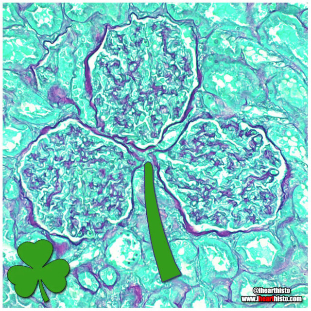
☘️Shamrock Gloms ☘️
For each petal on the shamrock,
This brings a wish your way.
Good health. Good luck. Happiness.
For today and every day.
Happy St. Patrick’s Day!
Three renal corpuscles (glomerulus + their surrounding Bowman’s capsules) floating in a sea of distal and proximal convoluted tubules within the cortex of the kidney.
These three small structures are knotted balls of capillaries (glomeruli) surrounded by a specialized epithelium (Bowman’s capsule) that is composed of cells called podocytes. These cells have tiny interlocking legs that form a small slit between them.
This structural organization is responsible for filtering your blood to produce a fluid that then travels within tubes continuous with the Bowman’s capsule called nephrons. In these nephrons the tubular fluid is modified by reabsorbing and secreting ions and conserving water to produce urine for excretion.

⚡Lord Voldermis⚡
A biopsy of a region of skin-that-shall-not-be-named (dermis/hypodermis junction shhh), complete with nerves, vessels, sweat glands and hair follicles.
by @nejiby

You can find LOVE in the strangest of places (2022 edition)
By row starting top left:
1. in a skin cylindroma
2. in an hepatic ductule
3. in a pancreas
4. in a warty penile growth
5. in a mucus-y colon
6. in a region of hypodermis
7. in a secondary oocyte
8. in a chondrosarcoma
9. in a small artery
Happy Valentine’s Day
Tag a friend with the histo heart you want to share with them and spread the love!
Images by:
@ihearthisto [1-4, 5-9]
@donna.horncastle [5]

Kitty Liver
Check meowt!
I’m a purrfect hepatic vein surrounded by hepatic lobules!
This is a hematoxylin and eosin stained slice through a liver that was injected with carbon/ink prior to fixation.
The liver is compose of numerous roughly hexagonal shaped lobules. Each lobule has a venule at its center (the central vein; top right) and a series of hepatic triads at its periphery. A triad is a collection of three structures - in this case branches of the hepatic artery, hepatic portal vein and hepatic duct.
Blood in the hepatic artery (oxygenated) and hepatic portal vein (rich in nutrients absorbed from the intestines) travels in sinusoids (the many narrow white spaces in this image) towards the central vein of each lobule. On its way through the sinusoids the blood is processed by the the many hepatocytes that line this region (the pink cells).
Within the sinusoids reside many liver macrophages (Kupffer cells) that phagocytose debris traveling through sinusoids. Normally these cells are invisible in standard H&E preparations but recall that this tissue was injected with ink! The ink was phagocytosed by the macrophages filling their cytoplasm with carbon so that they are now visible as black cells in the sinusoids.
Once the blood enters the central vein of each hepatic lobule they drain into the hepatic veins (kitty!) until the blood reaches the inferior venal cava.
In this way all blood, rich in raw nutrients and toxins absorbed from the GI tract is processed by the liver before it enters the systemic circulation!
Pawsitively unbeliverable!
Post link
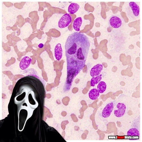
Ghostface Killer is back!
Only this time he’s hiding in a rectum!
Scaring while you’re learning!
A rectal scrape is performed (more often in veterinary med) to obtain a sample of the epithelial cells lining the rectum. These are observed in the microscope for abnormal changes or for microbes and their eggs/larvae that might be present in the digestive tract.
This particular scrape contains an epithelial cell that appears to be bi-nucleate and is surrounded by red blood cells.

Cookie Monster Ovary
Today’s ihearthisto is brought to you by the letter O for Oocyte!
This is image shows a high magnification view of a slice through an ovary.
You are looking at four tiny primordial follicles (the cookies above Cookie Monster’s head) located in the outer cortex of the ovary. Each of these follicles contains a single dormant, immature egg (a primary oocyte) that is halted in the the first phase of meiosis (prophase I). Eventually these follicles will be recruited to enter the maturation cycle that, against all the odds, could see them develop into an embryo.
Cookie monster himself is a larger multilaminar primary follicle (the cookie in his mouth is the nucleus of the oocyte within the follicle). This type of follicle has already been recruited and it’s follicular cells are dividing and differentiating. The oocyte within it though is still halted in prophase I of meiosis.
An oocyte will only complete meiosis if it is ovulated and fertilized by a spermatozoon (a sperm cell).
Post link
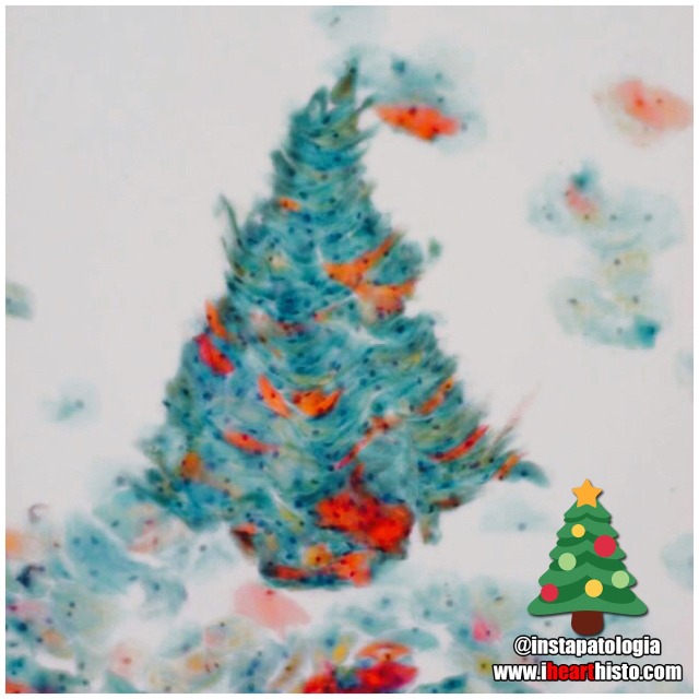
❄️Pappy Holidays❄️
There’s nothing quite like a Pap smear Christmas tree to rock around this happy holiday season!
by the awesome @instapatologia [Insta]

Have a Howelly-Jolly Christmas
A festive finding in the blood of an asplenic patient
A Howell–Jolly body is a cytopathological finding whereby small remnants of nuclear DNA are present in normally anuclear circulating erythrocytes.
During development in the bone marrow, late orthochromatophilic erythroblast normally expel their nuclei. However, in some cases, a small portion of DNA remains (the purple dots in the erythrocytes wearing the Santa hats).
Under normal circumstances if these irregular erythrocytes make it into the blood, they are removed from circulation by the spleen.
As a result, the presence of erythrocytes with Howell-Jolly bodies in peripheral blood smears like this usually signifies a damaged or absent spleen - because a healthy spleen would normally filter this type of red blood cell.
by exlibrisadpugno via reddit

Gingerbread Placenta
Run, run, run as fast as you can
You’ can’t catch me, I’m the chorionic villus gingerbread man!
The image shows a section through one of the many thousands of chorionic villi in the placenta that are responsible for the exchange of gas and nutrients with the maternal blood.
The mostly white space are the maternal blood lakes which are normally filled with mom’s blood while the small vessels (like gingerbread’s eyes and mouth) within the villus are branches of the umbilical vessels that shuttle blood back and forth to and from the growing baby.
The very thin cells lining the villus (gingerbread’s skin) are syncytiotrophoblast cells which gas must diffuse across in order to move from mom to baby and vice versa.
Histology by @BiopsyMD via Twitter

Rudolph the Red-Nosed Reindeer
Had a syncytiotrophoblast nose
And if you ever saw it
You would even say: “it forms the placental surface across which gases, nutrients & metabolites pass from the maternal circulation to enter the fetal circulation & vice versa”
This image shows many slices through the placental villi, fingerlike projections of the fetal-derived component of the human placenta. One of these villi looks like he’s been prohibited from participating in a variety of reindeer activities.
Rudolph’s core is composed of mesenchyme, an embryonic tissue that has the capacity to form tiny vessels (the tiny white holes) that are branches and tributaries of the umbilical arteries and vein that run in the umbilical cord and hook up with baby’s internal vascular plumbing.
The cell layer forming Rudolph’s skin (and his nose!) is called the syncytiotrophoblast layer. In the early placenta there are actually two layers (the outer syncytiotrophoblast and the inner cytotrophoblast). As the placenta matures the cytotrophoblasts thins and disappears leaving only the syncytiotrophoblast as the thin barrier between moms blood and those tiny fetal capillaries inside each villus.
But where is mom’s blood I hear you ask? Well, you can’t see it because it drained away once the placenta was removed and was sliced up. But… what you can see are the large ‘maternal blood lakes’ where her blood used to be (the large white spaces).
Now imagine all those fetal villi, and Rudolph, floating in those blood lakes and you can start to appreciate how efficient this arrangement is at allowing the exchange of gases and nutrients between mom and baby.

Prostate Snail
A secretory gastropod with a corpus amylaceum shell!
i❤️histo
This image is a close up view of a slice through the prostate gland!
The glandular portion of the prostate is composed of secretory acini that release prostatic fluid into a duct system within the gland. Prostatic fluid is a major component of semen and is rich in protein and sugar that keeps spermatozoa nourished as they travel through the reproductive tract.
The snail’s head and body are composed of the secretory epithelial cells!
The giant shell of the snail is a structure known as a prostatic concretion (corpus amylaceum or starchy body). This is a substance thought to be composed of thickened prostatic secretions and shed cells that is found in the acini and ducts of the prostate gland - it is of unknown significance.
However, these structures do increase in number with age and are a useful identifying feature of prostate in both non-pathological and pathological prostate specimens.
The space between the secretory acini is filled with a mixture of fibrous connective tissue and smooth muscle.
The smooth muscle is important because it contracts during ejaculation to push the prostatic fluid out of the gland and into the prostatic urethra where it mixes with spermatozoa arriving from the testis.
The connective tissue component is important clinically because as people with prostate glands grow older this fibrous tissue can undergo hyperplasia (excessive growth). This growth can constrict the urethra which passes directly through the prostate gland. The urethra, in addition to conveying semen during ejaculation, also carries urine during micturition/urination. This explains some of the symptoms associated with the common disorder, benign prostatic hyperplasia when urination becomes problematic for the elderly.
Post link
Students:
Histology:
- - - -
Those basic tissues are tough. But you have got this.

VERYSmooth Muscle
i♡histo

The Cytology Zoo
You can come too, too, too!
An exhibition of smears featuring some familiar looking squamous epithelial cell critters.
Can you recognize them all?
Cytology is the study of individual cells that have been isolated from a specimen. The technique is used mainly to study or screen for cancer but can be useful in the diagnosis of other diseases too, like those involving infectious organisms.
Most of the cells seen in a cytological smear or specimen are obtained in one of three ways:
- Scraping/Brushing the surface of a tissue (this is how cells are obtained from the uterine cervix during a pap smear)
- Collecting a fluid, this could be urine, mucus, blood or semen
- Fine-needle aspirations, this is when cells are removed by sucking them through a fine diameter needle. Fluid from the abdominal, pleural or pericardial cavities are obtained in this way and also cerebrospinal fluid during a lumbar puncture (or spinal tap).
The cells seen here are all squamous epithelial cells that are obtained from the cervix by scraping/brushing.
Credits:
@oreoimc (1-4) & (7-9)
@isac.p (5)
@sza_jhycto (6)
#histology #science #pathology #pathologists #anatomy #autopsy #zoo #animals #cytology #dentalschool #iquizhisto #premed #biology #medicaleducation #meded #nurse #nursing #medschool #medstudent #medicine #medlab #vetscience #vetschool #vetstudent #histologia #histotech #histo #pathArt #ihearthisto
![The Organ Rings [Answers] Original post without answers: https://www.ihearthisto.com/post/657148528](https://64.media.tumblr.com/484b98cf82c61d709722bca340d7cc7e/23e52e0dade3875d-bb/s1280x1920/3ce8479fe1e20400e90d61820fce13c514d6a592.jpg "The Organ Rings [Answers] Original post without answers: https://www.ihearthisto.com/post/657148528")
The Organ Rings [Answers]
Original post without answers:
https://www.ihearthisto.com/post/657148528765911040/the-organ-rings-to-commemorate-the-olympic
Trachea: C-shaped cartilage, a pseudostratified epi with cilia & goblet cells
Esophagus: Non-keratinized stratified squamous epi and muscularis mucosa
⚫ Artery (distributing): Prominent internal elastic lamina & thick smooth muscle tunica media
Bladder or Ureter: Transitional epithelium
Appendix: Crypts of Lieberkuhn (no villi) with surrounding submucosal lymph nodules
Thanks for playing!
Enjoy the games!
Post link

The Organ Rings
To commemorate the Olympic Games here are some slices through a few of the more tubular organs in your body!
Do you know what they are? Comment below!
Winner gets the gold medal!
[I’ll post the answers soon!]
Post link

Jurassic Pork
The pork tapeworm Taenia solium or a brachiosaurus?
i♡histo
This image shows the tapeworm T. solium, a parasite found in the bowels of humans who eat under-cooked pork.
You can see the proglottid segments of the worm’s neck/body and its terrifying scolex (head) complete with a hooked rostellum which it uses to attach itself onto your intestinal wall so that you don’t poop it out easily.
Fully grown worms can expand to around 2-3 meters in length and are composed of numerous segments each of which form their own reproductive unit.
The life cycle of the parasite begins when pigs ingest the T. solium eggs (from fecal contamination). The eggs develop into larvae inside the pig & then migrate through the intestinal wall to form cysts in their muscles & tissues.
Once slaughtered for meat, the larvae can be ingested by humans if the pork is eaten raw or under-cooked. Once in the human small intestine the larvae grow into large adult worms where they persist often unnoticed due to the lack of symptoms associated with their existence. The worms lay eggs into the intestines which are released during defecation thus continuing the life cycle.
“Life finds a way” - Dr. Ian Malcolm, Jurassic Park
A major complication of infection with T. solium is Cysticercosis. This is a parasitic tissue infection caused by the larval cysts of the tapeworm. These larval cysts infect brain, muscle, or other tissue, and are a cause of adult onset seizures. A person only gets cysticercosis by swallowing the eggs found in the feces of a person who has an intestinal tapeworm NOT from contracting a tapeworm by eating under-cooked pork itself. So people living in the same household with someone who has a tapeworm have a much higher risk of getting cysticercosis than people who don’t because they can be exposed to the eggs released in the stool.
Post link

Mi-fourth-sis
Dividing pyrotechnics!
Don’t forget to review the stages of mitosis while watching the fireworks this weekend!
It looks like this one is in metaphase!
Happy 4th of July!

❤️ Thanks Dad!
These are real spermatozoa.
Incidentally, based on head morphology, they are probably from a bull and not a human.
They were observed and photographed using phase contrast microscopy, a technique commonly used in semen analysis.
Note that the spermatozoa were realigned using photo editing software - spermatazoa are pretty cool cells but they do need a little help with their spelling sometimes.

SpongyBone SquarePants
Who lives in a bony medullary cavity?
Cancellous and yellow and porous is he.
Spongy bone!
Spongy bone is so named because its morphology (interconnected bony trabeculae/rods surrounding marrow spaces) resembles that of a sponge!
You can find spongy bone within flat bones (it forms the diplöe) and in long bones where it is located in the epiphyses (the ends of the bone) and diaphysis (the shaft of the bone).
Don’t be deceived by its name though. Spongy bone is not soft and spongy - it is still very strong, mature bone. It’s meshwork of trabeculae gives the inside of your bones strength and structure but at the same time, the space makes them more lightweight.
Spongy bone is always surrounded by a layer of compact bone (aka cortical bone).
Compact/cortical bone is different from spongy bone. It is named after the fact that it is more dense (i.e it isn’t made of rods and doesn’t have large spaces in it) and forms the outer aspect of a bone. If your bones were made entirely of compact bone than they would be much heavier and there would be no room for your bone marrow which is essential for making red and white blood cells.
Other names for spongy bone include ‘cancellous’ or 'trabecular’ bone.

A Pacinian Fingerprint at the Scene of the Crime
With all of this criminal activity it’s not surprising the body is full of cells!
A Pacinian or lamellar corpuscle is a mechanoreceptor that is most frequently found at the junction of the dermis/hypodermis in the skin. They are particularly prominent in the skin of the fingers and palms. In addition to the skin they are also found in other organs (e.g. mesentery, pancreas).
These very unique fingerprint-like structures have characteristic whorls formed from concentric lamella. Each lamella is separated from the next by a fluid filled space. And each lamella is lined on its inner and outer surface by connective tissue synthesized by the fibroblasts also in this region (you can see the nuclei of the fibroblasts in these lamellae in this image).
A nerve runs from the inner core of the corpuscle and is responsible for the transduction of mechanical forces applied to the skin into electrical signals that are conveyed to the central nervous system via the dorsal root ganglion.
This organization of fluid filled lamella and a central nerve are highly sensitive to pressure, particularly vibrational forces generated during tasks that involve a manipulative or tactile nature, like sensing the texture of a surface that is touched.
In fact, the screen of the phone/tablet you are using right now has been designed with the specific intention of stimulating the Pacinian corpuscles in your finger as you scroll.
So why not test out the Pacinians of your index finger for a moment by liking this post and following!! :-)
Post link

Mother Earth
A biopsy of the mammary gland obtained during pregnancy.
Let’s celebrate our planet and home!
Happy Earth Day everyone!
i♡histo
Post link

Easter Bunny Blood Cell
This monocyte is declaring a warren germs!
And doesn’t carrot all about what he phagocytoses.
He’ll just hop right to it.
Original source of histology is unknown

Which came first?
A seasonal conundrum in some keratin debris within a benign lymphoepithelial cyst.
Happy Spring & Happy Easter everyone!
The image shows a swirl of keratin debris (the chicken) in a small epithelial cell nest (the egg). The salivary gland is packed full of lymphocytes (the many, many purple nuclei surrounding the epithelial nest) which are a type of white blood cell.
Salivary gland lymphoepithelial cyst like this are rare and benign. Once the cyst is removed surgically from the gland it rarely recurs.

Dinosmear
A rare sighting of the rawrsome Papanicolaous Rex!
The cells in this image are the squamous epithelial cells that line the region of the ectocervix the region of the hole (os) in the cervix where it protrudes into the vagina.
Doctors obtain these cells by scraping the cervix. The cells are then smeared onto a slide and stained with the Papanicolaou stain during the pap smear. Cytologists examine the cells for any signs of abnormal morphology that could be an indicator of cervical cancer or other pathology.
Image based on the original by @mik__e [Insta]

⭐ Patrick Star in a Sperm Tube
My favorite Patrick in honor of #stpaddysday☘️
Some say that Patrick Star and the male reproductive tract are fairly similar… but if you look closely there’s actually a vas deferens.
This image shows the tube through which spermatozoa travel during ejaculation. It is called the vas deferens (ductus deferens).
Histologically it is unique because of its unique pseudostratified epithelium (surrounding Patrick…check out the unique regular beaded appearance of the basal cells in this epithelium) and its triple-layered smooth muscle wall - so thick you can actually palpate it within the spermatic cord within the scrotum. Patrick is actually within the empty space of the lumen of the tube (where the sperm travels).
Spermatozoa are produced in the seminiferous tubules of the testes and travel to the epididymis where they complete the maturation process. During ejaculation, a peristaltic wave of strong muscular contractions propel the sperm from the epididymis and into the vas deferens. This wave of peristalsis continues along the triple-thick muscle wall of the vas deferens eventually pushing the sperm out into the urethra. From here they have enough momentum to travel along the length of the penis before exiting exit the tube at the external urethral meatus (the fancy anatomical name for the hole at the end of the penis).
But sperm are not the only stuff that is ejaculated… at the same time as the sperm is moving along this pathway, the smooth muscle surrounding a number of glands also contracts which pushes their secretions into the urethra too at the same moment that the sperm is moving through it. These include secretions from the seminal vesicles (seminal fluid), prostate gland (prostatic fluid) and bulbourethral glands.
The final concoction that is ejaculated is actually a combination of sperm and all of these extra sperm-nourishing secretions and collectively it is known as semen.
Which brings us back to where we started with this sea-man - Patrick Star!
#histology #science #pathology #pathologists #anatomy #autopsy #patrickstar #spongebob #vasdeferens #malereproductive #stpatricksday #menshealth #premed #biology #medicaleducation #meded #nurse #nursing #medschool #medstudent #medicine #medlab #vetscience #vetschool #ihearthisto
Post link

The Ro-lung Stones
The Stones’ ‘Hot Lips’ logo formed from a blood vessel filled with erythrocytes within a congested zone around a focal pneumonia of the lung.
During the first stages of focal/lobar pneumonia macrophages (the larger cells that are visible in the white alveolar space) respond to phagocytose (eat) any pathogens in the lung.
Additionally, the small blood vessels within the lung tissue begin to engorge (you can see all the erythrocytes in the dilated vessels not only in the large 'hot lips’ vessel but the smaller vessels in the interalveolar septa between adjacent alveoli). This is the congestion phase.
With this dilation of vessels come more white blood cells, you can see numerous neutrophils (they are the small cells that look like they have multiple lobes to their purple nuclei) have migrated out of the vessels (a process called extravasation) into the surround tissue and alveolar spaces. These cells are signs of an additional immune response and they help the macrophages destroy pathogens in the airway.
Image is by @beautiful_pathologist - check out her Instagram for more histology.
Post link

Happy Vagina Scalp Epididymis Liver!!
Here’s to a healthier, happier New Year for everyone.
Image shows:
2 - Blood vessel in vaginal mucosa
0 - Hair follicle in scalp
2 - Epididymis
1 - Hepatic portal vein branch in liver
 in the digestive")
Santa Larvae
Hurry down my cecum tonight!
A nematode larva (Contracaecum) in the digestive tract of a seal. The eggs of this parasitic nematode use fish as an intermediate host before infecting piscivorous mammals, including humans who may forget to clean their holiday salmon!
This festive image was captured by veterinary pathologist @velphegor_vetpath via Instagram.
Happy holidays everyone!
Post link

A Charlie Brown Marrow:Happy Thanksgiving!
A developing leukocyte (white blood cell) observed in a sample of bone marrow obtained from the head of a femur
 looks like poo")
Blobfish-face
A coronal section through a developing jaw (when flipped upside down) looks like poor old blobfish!
The blobfish eyes are formed from Meckel’s cartilage. A component of the first pharyngeal arch that runs the length of the developing mandible. It degenerates as the fetus develops leaving only two small components on each side of the head. These ossify (become bone) to form the incus & malleus (ear ossicles) of the middle ear.
The blobfish nose is the developing tongue. It is composed of developing skeletal muscle fibers. Skeletal muscle forms from myoblasts that line up & fuse to form long myotubes. These will then synthesize actin/myosin which will allow them to contract & form the intrinsic skeletal muscle of the tongue.
The blobfish chin is formed by the developing maxilla. Two regions of tissue (the palatine shelves) grow together & fuse in the midline to form the posterior hard palate. You can see the midline suture forming & feel it in your own mouth by running your tongue along the roof of your mouth. Failure of these to fuse results in a variety of cleft lip and palate combinations.
The blobfish head is formed from a developing mandible. You can see small islands of bone forming within the mesenchymal tissue of the head. This type of bone development is called intramembranous ossification.
The blobfish is native to coastal waters off mainland Australia and Tasmania where it lives way deep down in the darkest depths of the ocean. Its gelatinous body is ideal for withstanding the pressure down there but when brought to the surface it looks like a sad melted pink crayon.
#histology #science #pathology #pathologists #anatomy #autopsy #blobfish #embryology #premed #biology #dentalschool #dentalstudent #dentistry #medicaleducation #meded #nurse #nursing #medschool #medstudent #medicine #medlab #vetscience #vetschool #vetstudent #histologia #histotech #histo #pathArt #ihearthisto
Post link

Caaaaarrrggghhh-diovascular Histology
Terrifying a medical student near you this Halloween!
The zombie’s lifeless eyes are arterioles.
His blood-filled mouth is a small vein.
All wrapped up in a connective tissue face.
Happy Halloween Histo fans!
Post link

The Creepiest Fibroblast You Will Ever See
With a nucleus straight from the fiery depths of hell*
*Note: Fibroblasts do not come from hell. They are actually derived from embryonic mesoderm and the creepiest thing they do is secrete the collagen and ground substance of connective tissue. Not very creepy at all really when you think about it.
Histology is from the microscope of the ‘fiancee of ned4spd8834’
Post link