#placenta


Gingerbread Placenta
Run, run, run as fast as you can
You’ can’t catch me, I’m the chorionic villus gingerbread man!
The image shows a section through one of the many thousands of chorionic villi in the placenta that are responsible for the exchange of gas and nutrients with the maternal blood.
The mostly white space are the maternal blood lakes which are normally filled with mom’s blood while the small vessels (like gingerbread’s eyes and mouth) within the villus are branches of the umbilical vessels that shuttle blood back and forth to and from the growing baby.
The very thin cells lining the villus (gingerbread’s skin) are syncytiotrophoblast cells which gas must diffuse across in order to move from mom to baby and vice versa.
Histology by @BiopsyMD via Twitter

Rudolph the Red-Nosed Reindeer
Had a syncytiotrophoblast nose
And if you ever saw it
You would even say: “it forms the placental surface across which gases, nutrients & metabolites pass from the maternal circulation to enter the fetal circulation & vice versa”
This image shows many slices through the placental villi, fingerlike projections of the fetal-derived component of the human placenta. One of these villi looks like he’s been prohibited from participating in a variety of reindeer activities.
Rudolph’s core is composed of mesenchyme, an embryonic tissue that has the capacity to form tiny vessels (the tiny white holes) that are branches and tributaries of the umbilical arteries and vein that run in the umbilical cord and hook up with baby’s internal vascular plumbing.
The cell layer forming Rudolph’s skin (and his nose!) is called the syncytiotrophoblast layer. In the early placenta there are actually two layers (the outer syncytiotrophoblast and the inner cytotrophoblast). As the placenta matures the cytotrophoblasts thins and disappears leaving only the syncytiotrophoblast as the thin barrier between moms blood and those tiny fetal capillaries inside each villus.
But where is mom’s blood I hear you ask? Well, you can’t see it because it drained away once the placenta was removed and was sliced up. But… what you can see are the large ‘maternal blood lakes’ where her blood used to be (the large white spaces).
Now imagine all those fetal villi, and Rudolph, floating in those blood lakes and you can start to appreciate how efficient this arrangement is at allowing the exchange of gases and nutrients between mom and baby.

Now that you’re 9 weeks pregnant, your baby is about the size of a fig. The heart has divided into four chambers, and the placenta is formed and ready to do its big work of producing specific hormones for your pregnancy. http://bit.ly/SDTFGB
Post link
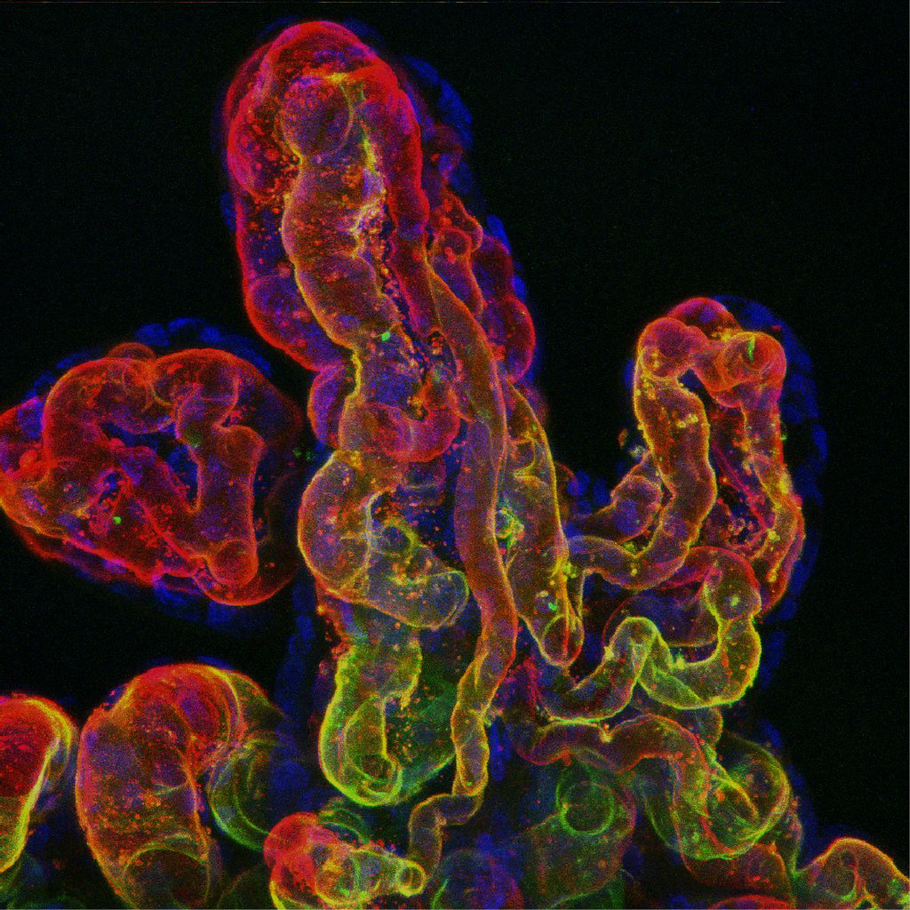
Romina Plitman Mayo: The feto-placental microvasculature by Department of Engineering, University of Cambridge
Via Flickr:
An attempt to label the different components of the fetal microvasculature in a perfused-fixed human placenta. The confocal microscopic image shows the fetal capillaries, where nutrients and waste is exchanged between a mother and her developing fetus. The complex architecture and biological system make the human placenta a unique organ and a challenge to investigate.


Moonpaintings this month :D Someone please push me to make a book, I think I have enough to make one now XD
Post link


Mom’s in control – even before you’re born
Researchers have uncovered a way in which information contained in unfertilised eggs influences the development of the fetus and placenta during pregnancy.
The research, performed in mice, indicates that even before conception a mother’s health may influence the health of her fetus.
Epigenetic information is critical for determining which genes are turned on and off in our DNA.
The researchers discovered that some epigenetic ‘marks’ laid down in eggs during their development in the ovaries and after fertilisation are passed onto the fetus and placenta.
The findings suggest that mothers have the genetic tools to control the growth of their offspring during pregnancy by instructing placental development.
Image credit: lunar caustic
Post link