#dental student


✨ Happy Star Wars Day ✨
May the Fourth be with you!
i♡histo
Star Wars histology from top left, clockwise:
1. The Graafian follicle Death Star
In a galaxy far, far away an intergalactic superweapon is halted in metaphase II of meiosis amid a surge in Luteinizing Hormone.
2. Jabba the Corpus Albicans
“makingsa lee ka bok pateesa… beeska chata wnow kong bantha poodoo”
(translation) “you may have been a good friend..but now you are bantha fodder”
The corpus albicans is a structure in the ovary that is formed when the corpus luteum regresses.
3. Tusken Raider in the Liver
Despite what you see here, Tusken Raiders are not native to the human liver. If you think that, then you are making a wookie mistake.
The image is actually a portal triad and demonstrates the major structures that enter and leave the liver: hepatic artery, hepatic portal vein and bile duct.
4. The Empire Strikes Back (at the Liver)
Liver histology is definitely where it’s At-At!
A region of connective tissue among the hepatocytes in the liver.
—
Images by @ihearthisto,@drjohnrajala,@zenonichand@hopkins_gi_pathrespectively
Post link
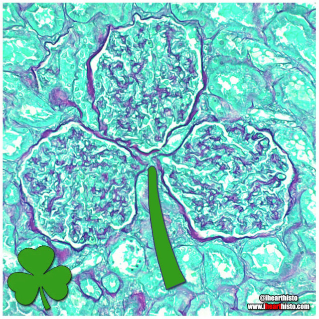
☘️Shamrock Gloms ☘️
For each petal on the shamrock,
This brings a wish your way.
Good health. Good luck. Happiness.
For today and every day.
Happy St. Patrick’s Day!
Three renal corpuscles (glomerulus + their surrounding Bowman’s capsules) floating in a sea of distal and proximal convoluted tubules within the cortex of the kidney.
These three small structures are knotted balls of capillaries (glomeruli) surrounded by a specialized epithelium (Bowman’s capsule) that is composed of cells called podocytes. These cells have tiny interlocking legs that form a small slit between them.
This structural organization is responsible for filtering your blood to produce a fluid that then travels within tubes continuous with the Bowman’s capsule called nephrons. In these nephrons the tubular fluid is modified by reabsorbing and secreting ions and conserving water to produce urine for excretion.

You can find LOVE in the strangest of places (2022 edition)
By row starting top left:
1. in a skin cylindroma
2. in an hepatic ductule
3. in a pancreas
4. in a warty penile growth
5. in a mucus-y colon
6. in a region of hypodermis
7. in a secondary oocyte
8. in a chondrosarcoma
9. in a small artery
Happy Valentine’s Day
Tag a friend with the histo heart you want to share with them and spread the love!
Images by:
@ihearthisto [1-4, 5-9]
@donna.horncastle [5]
Students:
Histology:
- - - -
Those basic tissues are tough. But you have got this.

VERYSmooth Muscle
i♡histo

The Organ Rings
To commemorate the Olympic Games here are some slices through a few of the more tubular organs in your body!
Do you know what they are? Comment below!
Winner gets the gold medal!
[I’ll post the answers soon!]
Post link

Mi-fourth-sis
Dividing pyrotechnics!
Don’t forget to review the stages of mitosis while watching the fireworks this weekend!
It looks like this one is in metaphase!
Happy 4th of July!

SpongyBone SquarePants
Who lives in a bony medullary cavity?
Cancellous and yellow and porous is he.
Spongy bone!
Spongy bone is so named because its morphology (interconnected bony trabeculae/rods surrounding marrow spaces) resembles that of a sponge!
You can find spongy bone within flat bones (it forms the diplöe) and in long bones where it is located in the epiphyses (the ends of the bone) and diaphysis (the shaft of the bone).
Don’t be deceived by its name though. Spongy bone is not soft and spongy - it is still very strong, mature bone. It’s meshwork of trabeculae gives the inside of your bones strength and structure but at the same time, the space makes them more lightweight.
Spongy bone is always surrounded by a layer of compact bone (aka cortical bone).
Compact/cortical bone is different from spongy bone. It is named after the fact that it is more dense (i.e it isn’t made of rods and doesn’t have large spaces in it) and forms the outer aspect of a bone. If your bones were made entirely of compact bone than they would be much heavier and there would be no room for your bone marrow which is essential for making red and white blood cells.
Other names for spongy bone include ‘cancellous’ or 'trabecular’ bone.

A Pacinian Fingerprint at the Scene of the Crime
With all of this criminal activity it’s not surprising the body is full of cells!
A Pacinian or lamellar corpuscle is a mechanoreceptor that is most frequently found at the junction of the dermis/hypodermis in the skin. They are particularly prominent in the skin of the fingers and palms. In addition to the skin they are also found in other organs (e.g. mesentery, pancreas).
These very unique fingerprint-like structures have characteristic whorls formed from concentric lamella. Each lamella is separated from the next by a fluid filled space. And each lamella is lined on its inner and outer surface by connective tissue synthesized by the fibroblasts also in this region (you can see the nuclei of the fibroblasts in these lamellae in this image).
A nerve runs from the inner core of the corpuscle and is responsible for the transduction of mechanical forces applied to the skin into electrical signals that are conveyed to the central nervous system via the dorsal root ganglion.
This organization of fluid filled lamella and a central nerve are highly sensitive to pressure, particularly vibrational forces generated during tasks that involve a manipulative or tactile nature, like sensing the texture of a surface that is touched.
In fact, the screen of the phone/tablet you are using right now has been designed with the specific intention of stimulating the Pacinian corpuscles in your finger as you scroll.
So why not test out the Pacinians of your index finger for a moment by liking this post and following!! :-)
Post link

Mother Earth
A biopsy of the mammary gland obtained during pregnancy.
Let’s celebrate our planet and home!
Happy Earth Day everyone!
i♡histo
Post link

Easter Bunny Blood Cell
This monocyte is declaring a warren germs!
And doesn’t carrot all about what he phagocytoses.
He’ll just hop right to it.
Original source of histology is unknown

Which came first?
A seasonal conundrum in some keratin debris within a benign lymphoepithelial cyst.
Happy Spring & Happy Easter everyone!
The image shows a swirl of keratin debris (the chicken) in a small epithelial cell nest (the egg). The salivary gland is packed full of lymphocytes (the many, many purple nuclei surrounding the epithelial nest) which are a type of white blood cell.
Salivary gland lymphoepithelial cyst like this are rare and benign. Once the cyst is removed surgically from the gland it rarely recurs.

The Ro-lung Stones
The Stones’ ‘Hot Lips’ logo formed from a blood vessel filled with erythrocytes within a congested zone around a focal pneumonia of the lung.
During the first stages of focal/lobar pneumonia macrophages (the larger cells that are visible in the white alveolar space) respond to phagocytose (eat) any pathogens in the lung.
Additionally, the small blood vessels within the lung tissue begin to engorge (you can see all the erythrocytes in the dilated vessels not only in the large 'hot lips’ vessel but the smaller vessels in the interalveolar septa between adjacent alveoli). This is the congestion phase.
With this dilation of vessels come more white blood cells, you can see numerous neutrophils (they are the small cells that look like they have multiple lobes to their purple nuclei) have migrated out of the vessels (a process called extravasation) into the surround tissue and alveolar spaces. These cells are signs of an additional immune response and they help the macrophages destroy pathogens in the airway.
Image is by @beautiful_pathologist - check out her Instagram for more histology.
Post link

A Charlie Brown Marrow:Happy Thanksgiving!
A developing leukocyte (white blood cell) observed in a sample of bone marrow obtained from the head of a femur
 looks like poo")
Blobfish-face
A coronal section through a developing jaw (when flipped upside down) looks like poor old blobfish!
The blobfish eyes are formed from Meckel’s cartilage. A component of the first pharyngeal arch that runs the length of the developing mandible. It degenerates as the fetus develops leaving only two small components on each side of the head. These ossify (become bone) to form the incus & malleus (ear ossicles) of the middle ear.
The blobfish nose is the developing tongue. It is composed of developing skeletal muscle fibers. Skeletal muscle forms from myoblasts that line up & fuse to form long myotubes. These will then synthesize actin/myosin which will allow them to contract & form the intrinsic skeletal muscle of the tongue.
The blobfish chin is formed by the developing maxilla. Two regions of tissue (the palatine shelves) grow together & fuse in the midline to form the posterior hard palate. You can see the midline suture forming & feel it in your own mouth by running your tongue along the roof of your mouth. Failure of these to fuse results in a variety of cleft lip and palate combinations.
The blobfish head is formed from a developing mandible. You can see small islands of bone forming within the mesenchymal tissue of the head. This type of bone development is called intramembranous ossification.
The blobfish is native to coastal waters off mainland Australia and Tasmania where it lives way deep down in the darkest depths of the ocean. Its gelatinous body is ideal for withstanding the pressure down there but when brought to the surface it looks like a sad melted pink crayon.
#histology #science #pathology #pathologists #anatomy #autopsy #blobfish #embryology #premed #biology #dentalschool #dentalstudent #dentistry #medicaleducation #meded #nurse #nursing #medschool #medstudent #medicine #medlab #vetscience #vetschool #vetstudent #histologia #histotech #histo #pathArt #ihearthisto
Post link

Caaaaarrrggghhh-diovascular Histology
Terrifying a medical student near you this Halloween!
The zombie’s lifeless eyes are arterioles.
His blood-filled mouth is a small vein.
All wrapped up in a connective tissue face.
Happy Halloween Histo fans!
Post link

The Creepiest Fibroblast You Will Ever See
With a nucleus straight from the fiery depths of hell*
*Note: Fibroblasts do not come from hell. They are actually derived from embryonic mesoderm and the creepiest thing they do is secrete the collagen and ground substance of connective tissue. Not very creepy at all really when you think about it.
Histology is from the microscope of the ‘fiancee of ned4spd8834’
Post link


6-23-17
OINA for life!
This is how i study OINA for head and neck muscles. I know it’s quite tedious but i’m a visual learner, memorizing it using tables is really not my thing. I just make tables for another summary, for example, a table showing the muscles that depresses the larynx, muscles innervated by ansa cervicalis and so on.
Post link