#medlab


Esoph-egg-us
✅Non-keratinized stratified squamous epithelium
✅Circumscribing muscularis mucosae
✅Submucosal glands
✅Dual layered muscularis externa
It’s just not Spring until you have painted your very own Esoph-egg-us
So get cracking and I don’t want to hear any eggscuses
#histology #science #pathology #pathologists #anatomy #autopsy #eggs #easter #spring #biology #esophagus #digestive #premed #meded #nurse #nursing #medschool #medstudent #medicine #education #vetscience #vetschool #dentistry #histotechnology #histologica #histotech #histo #pathArt #sciArt #ihearthisto
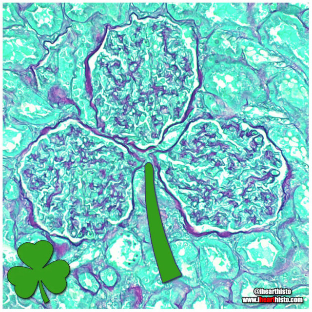
☘️Shamrock Gloms ☘️
For each petal on the shamrock,
This brings a wish your way.
Good health. Good luck. Happiness.
For today and every day.
Happy St. Patrick’s Day!
Three renal corpuscles (glomerulus + their surrounding Bowman’s capsules) floating in a sea of distal and proximal convoluted tubules within the cortex of the kidney.
These three small structures are knotted balls of capillaries (glomeruli) surrounded by a specialized epithelium (Bowman’s capsule) that is composed of cells called podocytes. These cells have tiny interlocking legs that form a small slit between them.
This structural organization is responsible for filtering your blood to produce a fluid that then travels within tubes continuous with the Bowman’s capsule called nephrons. In these nephrons the tubular fluid is modified by reabsorbing and secreting ions and conserving water to produce urine for excretion.

⚡Lord Voldermis⚡
A biopsy of a region of skin-that-shall-not-be-named (dermis/hypodermis junction shhh), complete with nerves, vessels, sweat glands and hair follicles.
by @nejiby
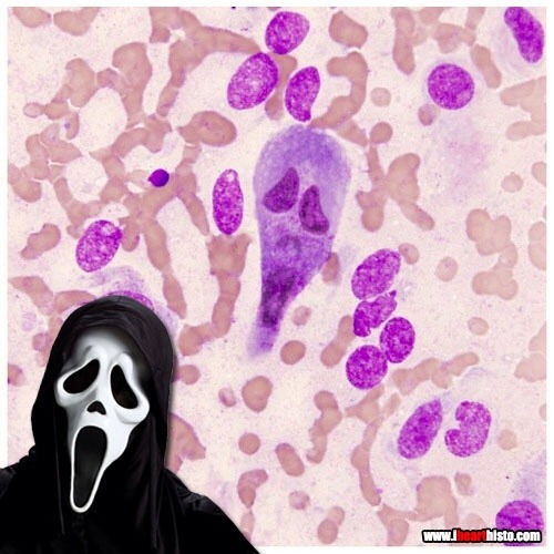
Ghostface Killer is back!
Only this time he’s hiding in a rectum!
Scaring while you’re learning!
A rectal scrape is performed (more often in veterinary med) to obtain a sample of the epithelial cells lining the rectum. These are observed in the microscope for abnormal changes or for microbes and their eggs/larvae that might be present in the digestive tract.
This particular scrape contains an epithelial cell that appears to be bi-nucleate and is surrounded by red blood cells.

Gingerbread Placenta
Run, run, run as fast as you can
You’ can’t catch me, I’m the chorionic villus gingerbread man!
The image shows a section through one of the many thousands of chorionic villi in the placenta that are responsible for the exchange of gas and nutrients with the maternal blood.
The mostly white space are the maternal blood lakes which are normally filled with mom’s blood while the small vessels (like gingerbread’s eyes and mouth) within the villus are branches of the umbilical vessels that shuttle blood back and forth to and from the growing baby.
The very thin cells lining the villus (gingerbread’s skin) are syncytiotrophoblast cells which gas must diffuse across in order to move from mom to baby and vice versa.
Histology by @BiopsyMD via Twitter

Rudolph the Red-Nosed Reindeer
Had a syncytiotrophoblast nose
And if you ever saw it
You would even say: “it forms the placental surface across which gases, nutrients & metabolites pass from the maternal circulation to enter the fetal circulation & vice versa”
This image shows many slices through the placental villi, fingerlike projections of the fetal-derived component of the human placenta. One of these villi looks like he’s been prohibited from participating in a variety of reindeer activities.
Rudolph’s core is composed of mesenchyme, an embryonic tissue that has the capacity to form tiny vessels (the tiny white holes) that are branches and tributaries of the umbilical arteries and vein that run in the umbilical cord and hook up with baby’s internal vascular plumbing.
The cell layer forming Rudolph’s skin (and his nose!) is called the syncytiotrophoblast layer. In the early placenta there are actually two layers (the outer syncytiotrophoblast and the inner cytotrophoblast). As the placenta matures the cytotrophoblasts thins and disappears leaving only the syncytiotrophoblast as the thin barrier between moms blood and those tiny fetal capillaries inside each villus.
But where is mom’s blood I hear you ask? Well, you can’t see it because it drained away once the placenta was removed and was sliced up. But… what you can see are the large ‘maternal blood lakes’ where her blood used to be (the large white spaces).
Now imagine all those fetal villi, and Rudolph, floating in those blood lakes and you can start to appreciate how efficient this arrangement is at allowing the exchange of gases and nutrients between mom and baby.
Students:
Histology:
- - - -
Those basic tissues are tough. But you have got this.

VERYSmooth Muscle
i♡histo

The Cytology Zoo
You can come too, too, too!
An exhibition of smears featuring some familiar looking squamous epithelial cell critters.
Can you recognize them all?
Cytology is the study of individual cells that have been isolated from a specimen. The technique is used mainly to study or screen for cancer but can be useful in the diagnosis of other diseases too, like those involving infectious organisms.
Most of the cells seen in a cytological smear or specimen are obtained in one of three ways:
- Scraping/Brushing the surface of a tissue (this is how cells are obtained from the uterine cervix during a pap smear)
- Collecting a fluid, this could be urine, mucus, blood or semen
- Fine-needle aspirations, this is when cells are removed by sucking them through a fine diameter needle. Fluid from the abdominal, pleural or pericardial cavities are obtained in this way and also cerebrospinal fluid during a lumbar puncture (or spinal tap).
The cells seen here are all squamous epithelial cells that are obtained from the cervix by scraping/brushing.
Credits:
@oreoimc (1-4) & (7-9)
@isac.p (5)
@sza_jhycto (6)
#histology #science #pathology #pathologists #anatomy #autopsy #zoo #animals #cytology #dentalschool #iquizhisto #premed #biology #medicaleducation #meded #nurse #nursing #medschool #medstudent #medicine #medlab #vetscience #vetschool #vetstudent #histologia #histotech #histo #pathArt #ihearthisto

❤️ Thanks Dad!
These are real spermatozoa.
Incidentally, based on head morphology, they are probably from a bull and not a human.
They were observed and photographed using phase contrast microscopy, a technique commonly used in semen analysis.
Note that the spermatozoa were realigned using photo editing software - spermatazoa are pretty cool cells but they do need a little help with their spelling sometimes.

SpongyBone SquarePants
Who lives in a bony medullary cavity?
Cancellous and yellow and porous is he.
Spongy bone!
Spongy bone is so named because its morphology (interconnected bony trabeculae/rods surrounding marrow spaces) resembles that of a sponge!
You can find spongy bone within flat bones (it forms the diplöe) and in long bones where it is located in the epiphyses (the ends of the bone) and diaphysis (the shaft of the bone).
Don’t be deceived by its name though. Spongy bone is not soft and spongy - it is still very strong, mature bone. It’s meshwork of trabeculae gives the inside of your bones strength and structure but at the same time, the space makes them more lightweight.
Spongy bone is always surrounded by a layer of compact bone (aka cortical bone).
Compact/cortical bone is different from spongy bone. It is named after the fact that it is more dense (i.e it isn’t made of rods and doesn’t have large spaces in it) and forms the outer aspect of a bone. If your bones were made entirely of compact bone than they would be much heavier and there would be no room for your bone marrow which is essential for making red and white blood cells.
Other names for spongy bone include ‘cancellous’ or 'trabecular’ bone.

Zomb-hair
A very close-up scene from the hit TV series ‘The Walking Dreads’
This is actually a vibrissae hair follicle from the face of a (a whisker).
Vibrissae are sensory hairs that are different from regular body hairs in the fact that they are surrounded by a blood filled sinus (z’s brain) and are associated with sensory neurons that have a distinct and representative pattern in the somatosensory cortex of the mouse brain. The fact that they are well mapped in the brain illustrates their importance in everyday behavior and survival - they are involved in things like detecting, orienting and sensing length of surfaces/objects and tracking (e.g. finding gaps in a maze and answering the question 'can my head fit through here?’).
Note also the sebaceous glands (z’s eyes) which secrete a lipid rich secretion called sebum for maintaining hair and epidermis keratin (keratin is the wispy stuff lining the skin at the bottom of the image and forming z’s lil arms).
The hair in this follicle is absent as you can see z’s wailing mouth is completely empty (where you might expect to see the shaft of a hair).
The closest things us humans have to vibrissae are the thick long hairs in our nostrils which are fairly sensitive to tickles (and make your eyes water if plucked) but more importantly provide a first line of filtration to prevent big particles from being inhaled into your nasal cavity.
#histology #science #anatomy #pathology #autopsy #pathologists #dermatology #halloween #hair #skin #zombie #dentalstudent #dentalschool #histotech #medlab #premed #meded #nurse #nursing #medschool #medstudent #medicine #vetscience #vetschool #histotechnology #histologia #histo #pathArt #vetstudent #ihearthisto
Post link