#immunofluorescence

New Shirt design up in my shop at moocyte.com!
Made using my own immunofluorescence microscopy images ♥️

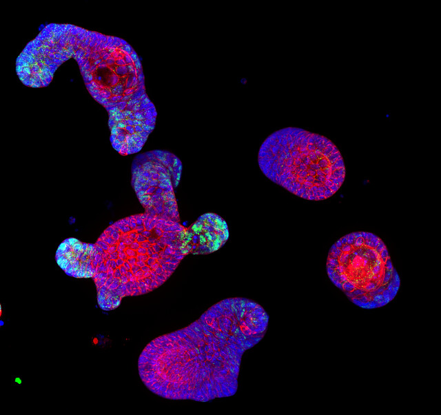
“Confocal immunofluorescence image of intestinal organoid” by Denise Serra, University of Basel by SNSF Scientific Image Competition
Via Flickr:
Entry in category 1. Object of study; © CC-BY-NC-ND: Denise Serra This image shows immuno-staining of mature intestinal organoids, complex 3-D structures that resemble both morphologically and functionally the intestinal epithelium. Intestinal organoids are great model system to study how the epithelium develops, regenerates after injury and maintain its physiological state. The image has been taken with a confocal microscope, in blue are nuclei (DAPI), in red a membrane protein and in green a transcription factor.
 Immunofluorescence image of endogenous epsin 1 (red")
Intracellular distribution of epsin 1. (A Upper) Immunofluorescence image of endogenous epsin 1 (red) and endogenous clathrin (green). (Lower) Magnified view of the boxed region. Shown are the epsin 1(Left), clathrin (Center), and overlaid (Right) images. (B Upper) Overlay of the Venus fluorescence image of epsin 1-Venus (green) and the immunofluorescence image of endogenous clathrin (red). The cell was transiently transfected with epsin 1-Venus. (Lower) Magnified view of the boxed region. Shown are the epsin 1-Venus (Left), clathrin (Center), and overlaid (Right) images. (Scale bars,10 μm.)
Chen Chen and Xiaowei Zhuang; Epsin 1 is a cargo-specific adaptor for the clathrin-mediated endocytosis of the influenza virus; PNAS August 19, 2008 105 (33) 11790-11795; https://doi.org/10.1073/pnas.0803711105
Post link
 and #nuclei using DAPI (green). T")
#mitochondria stained using #immunofluorescence for TOM20 (orange) and #nuclei using DAPI (green). To show the morphology of MEF cells, they used bright field #microscopy (detection with TPMT) . For confocal #imaging they utilized a @zeiss_micro LSM 710 at the LMF @DZNE_en.
Image courtesy of Christian Lamberg, PhD (@Christi23003438 on twitter) (see original post)
Post link

Colony of stem cells derived from an individual with bipolar disorder
Source : medicine.umich.edu
Post link
