#fluorescence microscopy

New Shirt design up in my shop at moocyte.com!
Made using my own immunofluorescence microscopy images ♥️

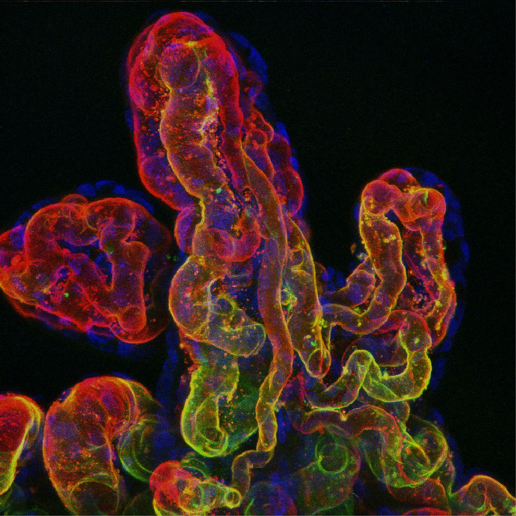
Romina Plitman Mayo: The feto-placental microvasculature by Department of Engineering, University of Cambridge
Via Flickr:
An attempt to label the different components of the fetal microvasculature in a perfused-fixed human placenta. The confocal microscopic image shows the fetal capillaries, where nutrients and waste is exchanged between a mother and her developing fetus. The complex architecture and biological system make the human placenta a unique organ and a challenge to investigate.
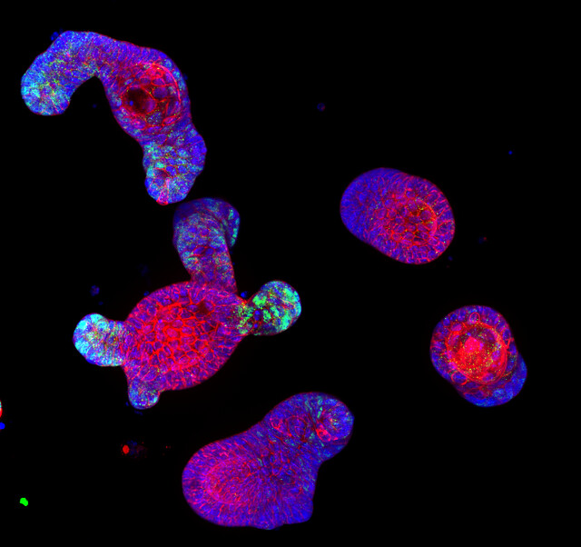
“Confocal immunofluorescence image of intestinal organoid” by Denise Serra, University of Basel by SNSF Scientific Image Competition
Via Flickr:
Entry in category 1. Object of study; © CC-BY-NC-ND: Denise Serra This image shows immuno-staining of mature intestinal organoids, complex 3-D structures that resemble both morphologically and functionally the intestinal epithelium. Intestinal organoids are great model system to study how the epithelium develops, regenerates after injury and maintain its physiological state. The image has been taken with a confocal microscope, in blue are nuclei (DAPI), in red a membrane protein and in green a transcription factor.
 Immunofluorescence image of endogenous epsin 1 (red")
Intracellular distribution of epsin 1. (A Upper) Immunofluorescence image of endogenous epsin 1 (red) and endogenous clathrin (green). (Lower) Magnified view of the boxed region. Shown are the epsin 1(Left), clathrin (Center), and overlaid (Right) images. (B Upper) Overlay of the Venus fluorescence image of epsin 1-Venus (green) and the immunofluorescence image of endogenous clathrin (red). The cell was transiently transfected with epsin 1-Venus. (Lower) Magnified view of the boxed region. Shown are the epsin 1-Venus (Left), clathrin (Center), and overlaid (Right) images. (Scale bars,10 μm.)
Chen Chen and Xiaowei Zhuang; Epsin 1 is a cargo-specific adaptor for the clathrin-mediated endocytosis of the influenza virus; PNAS August 19, 2008 105 (33) 11790-11795; https://doi.org/10.1073/pnas.0803711105
Post link

Colony of stem cells derived from an individual with bipolar disorder
Source : medicine.umich.edu
Post link

Stem cells in the skin - stem cells labelled in red are found in special microenvironments where they are surrounded by other types of cell.
Post link

A rendered image of a primary neuronal stem cell culture in which cells were labeled with different fluorescently labeled proteins that differentiate between stem cells (orange/yellow) and their neuronal ‘offspring’ (blue/ green/ purple).
Post link
, vinc")
Huntington’s stem cell derived oligodendrocyte precursors stained for phalloidin (green), vinculin (red) and DNA (blue).
Post link
 and Nissl bodies (green).")
Rat dorsal root ganglia cells stained for Alpha Acetyl tubulin (red) and Nissl bodies (green).
Post link
, GAD65 (green) and DNA (blue).")
Trisomy 21 derived neural cells stained for neurofilament heavy (red), GAD65 (green) and DNA (blue).
Post link

Mouse diaphragm consisting of neurons, muscle cells and neuromuscular junctions under the confocal microscope: Green structures show neurons (Alexa 488), red areas are neuromuscular junctions (rhodamin), and blues areas are muscle fiber (myosin, DODT contrast).
Post link

Human epithelial cell in mitosis, fluorescently labeled for alpha tubulin, gamma tubulin and DNA.
Post link

Image shows adult human fibroblast cells with intracellular proteins involved in adhesion of these cells to an extracellular matrix. Magenta represents actin stress fibers in a cell and green staining represents a focal adhesion protein vinculin, which together contribute to how strongly these cells adhere to a matrix surface. Blue is the nucleus of a cell.
Post link


Leishmania major promastiotes during infection of primary fibroblast culture. Cells are stained with anti-tubulin (green) and anti-actin (red).
Post link

 and DAPI (b")
Pilidium larva of the nemertean Cerebratulus sp.
Labelled with phalloidin (green: actin) and DAPI (blue: nuclei).
Post link

This microscopic fluorescent image of the mosquito’s circulatory system shows muscle cells in green; cellular DNA in blue and periostal hemocytes in orange.
Post link

Confocal micrograph of a blind cavefish embryo at around five days post-fertilisation viewed from the side (lateral view) with an antibody that targets a calcium binding protein (calretinin) shown in green, which highlights different neuronal types and their processes in the nervous system. The cavefish Mexican tetra (Astyanax mexicanus) has a seeing and a blind form; the latter lives in dark environments, and relies on other senses. The blind cavefish has specially adapted traits that its sighted relation (dwelling near the surface) does not. These include a greater number of sensory receptors and taste buds along its body; these taste buds are also more efficient than the equivalent cells in the seeing cavefish. The eyes are still present at this stage of development but they will degenerate naturally during the lifetime of the fish as they live in a dark environment where eyes are redundant. Adult cavefish are blind.
Post link
 covering the tail of a 3- day-old larval zebrafish. This")
Sensory axons (long, slender nerve fibers) covering the tail of a 3- day-old larval zebrafish. This “Brainbow” image was collected using confocal microscopy. In the Brainbow technique (Nature, 2007), cells randomly choose combinations of red, yellow and cyan fluorescent proteins, so that they each glow a particular color. This provides a way to distinguish neighboring cells of the nervous system and follow their pathways. Seventh Prize, 2009 Olympus BioScapes Digital Imaging Competition
Post link

L. polyphemus, Cast skin of a horseshoe crab Cuticular receptors Autofluorescence, Ex 488, Det 500-600 nm, Color Coded Projection Dr. Manfred Schmidt University of Hamburg, Zoological Institute, Neurophysiology, Martin-Luther-King-Platz 3, 20146 Hamburg
Post link