#neurobiology
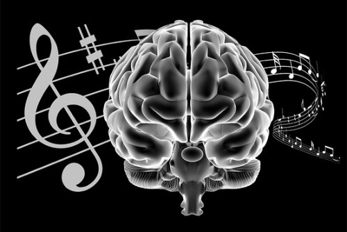
Community music programs enhance brain function in at-risk children
A Northwestern University study provides the first direct evidence that a community music program for at-risk youth has a biological effect on children’s developing nervous systems.
Two years of music lessons improved the precision with which the children’s brains distinguished similar speech sounds, a neural process that is linked to language and reading skills. One year of training, however, was insufficient to spark changes in the nervous system.
“This research demonstrates that community music programs can literally ‘remodel’ children’s brains in a way that improves sound processing, which could lead to better learning and language skills,” said study lead author Nina Kraus, the Hugh Knowles professor of communication sciences in the School of Communication and of neurobiology and physiology in the Weinberg College of Arts and Sciences at Northwestern.
“Music Enrichment Programs Improve the Neural Encoding of Speech in At-Risk Children,” published in the Journal of Neuroscience, is one of the few studies to evaluate biological changes following participation in an existing, successful music education program.
Post link
When the brain switches from deep non-REM sleep over to REM sleep, something remarkable happens: the brain erupts with spikes of activity in the MRI scans.
Specifically, 4 areas of the brain fire up when dreaming starts during REM sleep: the visuospatial regions, the motor cortex, the hippocampus, and the amygdala. In contrast to all of these areas, one part of the brain does the opposite. The left and right sides of your prefrontal cortex becomes markedly deactivated during REM sleep. This is important because your prefrontal cortex controls logical reasoning. This is, in part, why dreams are often filled with movement, strong emotions, past memories, people, and experiences, yet are utterly irrational.
When we are in REM sleep dreaming, the body is paralyzed, preventing us from acting out our bizarre dreams. Otherwise, we would put ourselves in danger and be popped out of the gene pool rather quickly!
A breakthrough neuromodulation system rapidly restores motor function in patients with a severe spinal cord injury (SCI), new research shows.
The study demonstrated that an epidural electrical stimulation (EES) system developed specifically for spinal cord injuries enabled three men with complete paralysis to stand, walk, cycle, swim, and move their torso within 1 day.
SCIs involve severed connections between the brain and extremities. To compensate for these lost connections, researchers have investigated stem cell therapy, brain-machine interfaces, and powered exoskeletons.
However, these approaches aren’t yet ready for prime time.
A rendered image of a primary neuronal stem cell culture in which cells were labeled with different fluorescently labeled proteins that differentiate between stem cells (orange/yellow) and their neuronal ‘offspring’ (blue/ green/ purple).
Post link
Trisomy 21 derived neural cells stained for neurofilament heavy (red), GAD65 (green) and DNA (blue).
Post link
Mouse diaphragm consisting of neurons, muscle cells and neuromuscular junctions under the confocal microscope: Green structures show neurons (Alexa 488), red areas are neuromuscular junctions (rhodamin), and blues areas are muscle fiber (myosin, DODT contrast).
Post link
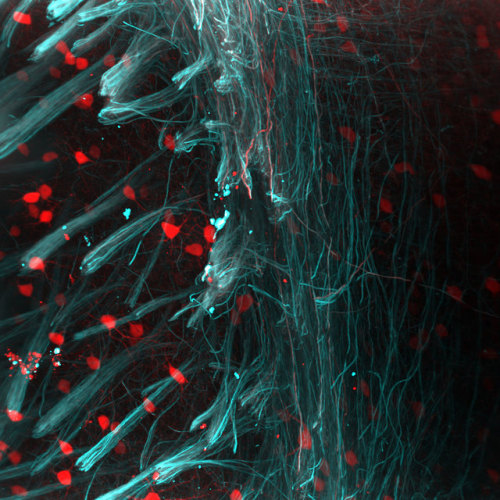
Sensory axons (long, slender nerve fibers) covering the tail of a 3- day-old larval zebrafish. This “Brainbow” image was collected using confocal microscopy. In the Brainbow technique (Nature, 2007), cells randomly choose combinations of red, yellow and cyan fluorescent proteins, so that they each glow a particular color. This provides a way to distinguish neighboring cells of the nervous system and follow their pathways. Seventh Prize, 2009 Olympus BioScapes Digital Imaging Competition
Post link
Release of Dopamine in Infant Brains May Help Control Early Social Development
Increased levels of dopamine release in the basolateral amygdala as a result of stressful situations during infancy could lead to lasting behavioral issues and social difficulties, a new study reports.
Post link
If we were able to detect consciousness and understand the process we could use it to first make neuronal networks with the process applied and then in super computers with the computational mass similar to the computational mass of a human brain - enabling the mind transfer.
If the super computers had downloaded the memories of the person and used the neuronal process of consciousness (within itself) we would probably be able to talk with the person as we knew them before the transfer (as who we are is actually a bunch of memories connected by the conviction of one continued and steady self).
But the problem might be - it wouldn’t be us. We, the consciousness that reads it, would no longer exist. This consciousness would not experience the transfer. It would be replaced with a new one. Any other person wouldn’t see the difference (from their perspective there would be none) - we would behave exactly like we did before as we would have same memories and experiences just managed by another consciousness (that would interpret them exactly as the old one).
It’s like with teleportation. To teleport you need to be destroyed in one place and rebuild in another. You would be exactly the same just made of different particles. Should it still be you? In some context yes.
Our first topic for you, as requested by our readers, is the biology of brains!
Tell us everything you know about brains– how they’re structured, and how they work. A few questions to get you thinking:
- What’s the history of the biology of brains?
- How are brains organized?
- How do brains function?
- How do brains differ across species?
- Where do you think science is headed with regard to the biology of brains?
Note that all of these questions can be answered at many different levels and in thousands of different ways, not to mention that we encourage thinking outside of these guiding questions! We’re just asking you to give us a tiny bit of anything you know.
Again, the rules, for those new to our game:
- Reblog and add anythingthat comes to mind regarding the topic– facts you know, things you learned in school, questions you have, links to an article, papers you’ve read– we mean anything!
- If you don’t think you have anything to contribute, just reblog and pass the thread along to your followers.
That’s it! SciNote will be reading every single one of your responses, and given enough responses, we will combine your contributions, citing your work, in one big comprehensive article to be posted on SciNote.org.
Gene Therapy Reverses Effects of Autism-Linked Mutation in Brain Organoids
In a study published May 02, 2022 in Nature Communications, scientists at University of California San Diego School of Medicine used lab-grown human brain organoids to learn how a genetic mutation associated with autism disrupts neural development. Recovering the function of this single gene using gene therapy tools was effective in rescuing neural structure and function.
Autism spectrum disorders (ASD) and schizophrenia have been linked to mutations in Transcription Factor 4 (TCF4), an essential gene in brain development. Transcription factors regulate when other genes are turned on or off, so their presence, or lack thereof, can have a domino effect in the developing embryo. Still, little is known about what happens to the human brain when TCF4 is mutated.
To explore this question, the research team focused on Pitt-Hopkins Syndrome, an ASD specifically caused by mutations in TCF4. Children with the genetic condition have profound cognitive and motor disabilities and are typically non-verbal.
Using stem cell technology, the researchers created brain organoids, or “mini-brains,” using cells from Pitt-Hopkins Syndrome patients, and compared their neurodevelopment to controls.
They found that fewer neurons were produced in the TCF4-mutated organoids, and these cells were less excitable than normal. They also often remained clustered together instead of arranging themselves into finely-tuned neural circuits. This atypical cellular architecture disrupted the flow of neural activity in the mutated brain organoid, which authors said would likely contribute to impaired cognitive and motor function down the line.
The team thus tested two different gene therapy strategies for recovering the functional TCF4 gene in brain tissue. Both methods effectively increased TCF4 levels, and in doing so, corrected Pitt-Hopkins Syndrome phenotypes at molecular, cellular and electrophysiological scales.
“The fact that we can correct this one gene and the entire neural system reestablishes itself, even at a functional level, is amazing,” said senior study author Alysson R. Muotri, PhD, professor at UC San Diego School of Medicine.
The team is currently optimizing their recently licensed gene therapy tools in preparation for future clinical trials, in which spinal injections of a genetic vector would hopefully recover TCF4 function in the brain.
— Nicole Mlynaryk
Post link


