Can anyone help me understand it? I am not able to understand why +/- signs are written above the gel profile? And can anyone help me with the hint to the solution?
Here is a photograph of my current classmates and I DNA before undergoing gel electrophoresis.
The DNA was loaded into the wells (tiny rectangles) with pipettes. When the gel is electrically charged, the DNA travels from the negative terminal (the black circle at the top left) toward the positively charged anode (where the red circle is at the bottom right corner).
(Note: The three wells (green rectangles) of darkest color on the second row are mine, my DNA is on the first two, the last and third well has extra water. It is my negative response well, to test that there is no contamination. If there is any contamination it will be observed in that third well.)
Contamination refers to DNA of other things (animals, plants, etc) showing up instead of the intended DNA for which results are being analyzed.
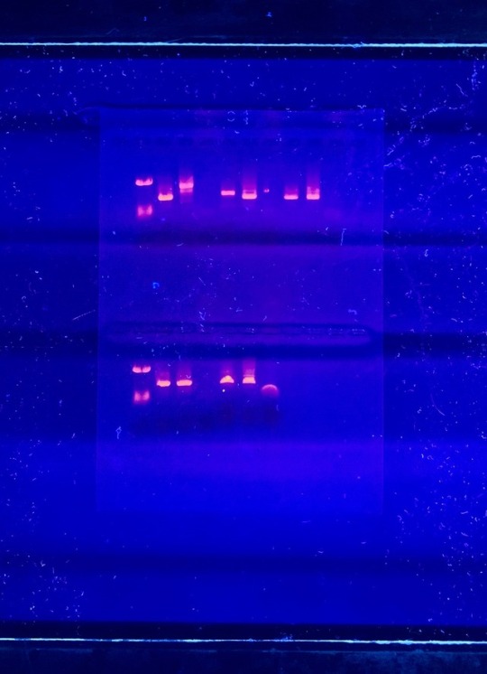
Here is the after photo. The DNA being viewed (since it was stained) under a UV light.
(Note: Another classmate added samples in after the first photo was taken. My DNA is in the second well from the left on the second row, and my positive is to the right of that well. The blank well in the center of the second row is my negative, not much showed which means there was little to no contamination in my DNA sample).
The gel used is made of agarose, which is easy to cast, and has relatively fewer charged groups. The agarose gel is suitable for separating DNA (and macromolecules) by size as it is charged (with electricity) and it travels through the pores in the gel (See some of the wells on the second image look more elongated).
DNA fragments can easily be extracted from the gel, which is a suitable electrophoresis buffer.
What are the applications of gel electrophoresis?
It can be used to estimate the size of DNA molecules.
To analyze PCR products, such as molecular genetic diagnosis, or genetic fingerprinting.
Joan A. Steitz (b. 1941) is a biologist whose research focused on RNA and its interaction with ribosomes, for which she won the Lasker-Koshland Award for Special Achievement in Medical Science.
She obtained her PhD from Harvard and conducted postdoctoral research at the University of Cambridge. Her research offered groundbreaking insight into autoimmune diseases, their causes and treatments. She is a Professor of Molecular Biophysics and Biochemistry at Yale University.
Gardening was my first introduction to plants. I enjoyed growing plants so much that I decided to study them. Or rather, I studied the growing of them,i.e.horticulture. During my studies, I became increasingly interested in botany, a vast scientific field that investigates all things plant related, from their evolutionary history to their cellular biology to their interactions with other…
Get to know Patricia Bunda, biomedical scientist and cancer researcher
1) What do you do?
My current research project focuses on creating mouse models of organ-specific cancer metastasis which are useful tools in pre-clinical testing of novel immunotherapy treatments and in studying the ability of cancer cells to preferentially metastasize to organs such as the brain and the bone.
Most, if not all, of us know someone who is suffering or had suffered from cancer. Being able to work on something that can help cure this disease is a unique privilege that motivates me on a daily basis.
2) Where do you work?
I currently work as a post-baccalaureate researcher at the National Cancer Institute, National Institutes of Health (NIH) in Bethesda, Maryland. Working in the largest biomedical facility in the world enables me to collaborate with experts and see the potential impact of my basic science research in the clinical setting.
3) Tell us about the photos!
[Top:] I was presenting the results of my senior thesis to one of the judges at the UCLA Undergraduate Research Poster Day. I remember being quite nervous but, at the same time, excited to present the results of my year-long project. Winning the Dean’s Prize was just icing on the cake!
[Bottom:] Apart from my research involvement, I also served as the medical outreach director for Pilipinos for Community Health (PCH) at UCLA during my senior year in college. I was in charge of planning and fundraising for our medical mission to the Philippines. This photo was taken at Sitio Kidadayaig in General Nakar, Quezon Province where our team helped provide basic medical services to over a hundred residents of this remote mountain community.
4) Tell us about your academic career path so far.
I graduated from Imus Institute Science High School in Imus, Cavite. My biology teacher, Mr. Donnie Legaspi, inspired me to study Biology in UP Los Baños. After only my sophomore year at UPLB, my family decided to immigrate to the US. I continued to pursue Biology at Palomar College and eventually transferred to University of California, Los Angeles (UCLA) where I graduated Summa Cum Laude with a Bachelor’s degree in Molecular, Cell, and Developmental Biology, minor in Biomedical Research. I am concurrently doing research and applying to MD/PhD programs here in the US.
5) Anything else you’d like to share?
According to a University of the Philippines study, 6 out of 10 Filipinos die without seeing a doctor. Growing up in a small town in the Philippines and seeing firsthand the reality of this statistic motivated me to pursue a career in medicine. However, my research experience in the US allowed me to realize that a good healthcare system and a strong research community go hand in hand. Every year, countless doctors, nurses, and scientists leave the Philippines in search of better employment opportunities overseas. This creates a shortage of high-skilled workers that are supposed to be integral parts of our country’s healthcare sector. I believe that promoting biomedical research is a powerful way to improve healthcare in the as it fosters a society that supports and encourages intellectuals to stay in their own country instead of seeking jobs abroad.
In 2013, the Philippines only had 187.7 scientists per million people, one of the lowest densities of researchers in the world. To all Pinoy Scientists out there, I urge you to advocate for and help develop the research field in our country. You can start small by mentoring an aspiring young pinoy scientist or go big and take advantage of the DOST Balik Scientist Program (https://bspms.dost.gov.ph/).
With the innate curiosity and resourcefulness of Filipinos, I believe that we have the potential to make a significant impact in the larger scientific community and help improve the lives of our countrymen. Mabuhay ang mga siyentipikong Pinoy!
P.S. I am planning to start a facebook group for current and aspiring Pinoy scientists to share experiences, ideas, and opportunities with each other. If you’re interested in helping me start this group, shoot me a message (facebook.com/patbunda). Also, feel free to contact me if you want to learn more about cancer research!
MeetChristine Jewel Yabut, molecular biologist and phylogenist
1) What do you do?
My research focuses on the Phylogeny of an insect group (Trichoptera) that can be found in clean streams and rivers. I discover and describe new species from my group. The species that I discovered from the Philippines were collected in the streams of Los Banos, Laguna inside the University of the Philippines - Los Banos (Diplectrona lagunensis)and in Imugan, Nueva Vizcaya (Hydropsyche philippinensis). During my collection in Imugan, I also collected another new species that was described by a colleague who named the species after me. Cheumatopsyche christinae! I am now finishing a barcode library of the Insect species from the group Trichoptera in South Korea.
2) Where do you work?
I just finished my Ph.D. in Molecular Biology in Korea University last August.
3) Tell us about the photos!
[Left:]This picture was taken in South Carolina, USA when I attended a training for Aquatic Insect collection. I did a teaching assistantship during the period of the summer class.
[Right:]This picture was from Mindanao when I did insect collection from January 21-25, 2014. These amazing local people helped me during my sampling.
4) Tell us about your academic career path so far.
I graduated with a degree in B.S. Biology at the University of the Philippines Baguio. Then I entered Korea University for an integrated M.S. and Ph.D. program. I am now searching for a postdoc position and plan to describe many new species from the Philippines.
5) Anything else you’d like to share?
I am now finishing a barcode library of Korean Insects called caddisfly. This barcode library is like a tag of DNA consisting of 658 basepairs of DNA nucleotide sequences that can be referred to identify the name of the species.
Name Alice Kim Location Prague, Czech Republic What do you do? I am an aspiring makeup artist, but I’m currently studying molecular biology at the Charles University. Honestly I’m at the beginning of my journey, so I haven’t had any struggles or any significant accomplishments yet. MoreTwitter|Instagram Contact[email protected] Share what you do. http://bit.ly/ADEsubmit.
The surfaces of all cells in nature are festooned with a complex and diverse array of sugar chains (called glycans). These perform a wide variety of biological functions, from the proper folding of proteins to cell-to-cell interactions. Their ubiquity in nature underscores their essentialness to complex life.
This week, the fourth edition of “Essentials of Glycobiology” (the study of glycans) was published by Cold Spring Harbor Laboratory Press. It’s a continuation and updating of landmark work by a consortium of editors, led by Ajit Varki, MD, Distinguished Professor in the departments of Medicine and Cellular and Molecular Medicine at UC San Diego School of Medicine, with contributions from a number of UC San Diego scientists and physicians, including Jeffrey D. Esko, PhD, Distinguished Professor of cellular and molecular medicine; Pascal Gagneux, PhD, professor of pathology and anthropology, and Kamil Godula, PhD, associate professor of chemistry and biochemistry, and Amanda Lewis PhD, professor of obstetrics-gynecology and reproductive science.
Varki and Esko are also founding directors of the Glycobiology Research and Training Center (GRTC) at UC San Diego, established in 1999, and have recently handed over leadership to Lewis and Godula.
Glycobiology is a relatively new scientific discipline. The term was only coined in 1988, recognizing the combining of carbohydrate chemistry and biochemistry to focus on glycans, which have since proven to have a multitude of diverse and often critical roles in biology.
They have been linked to human origins and as a key evolutionary marker. They are found to both inhibit and promote tumor growth; and the presence of a particular sialic acid in red meat may be linked to increased cancer risk in humans. Another class of glycans called glycosaminoglycans have been shown by Esko and colleagues to be involved in COVID-19 coronavirus pathogenesis. The cover of the fourth edition presents an all-atom model of infamous spike protein of the pandemic virus, emphasizing the massive array of glycan chains modelled by UC San Diego professor of biology Rommie Amaro.
Varki, Esko and colleagues at the GRTC have been central to many of the advances in glycobiology, and the textbook, which originally debuted in 1999, has been an enduring effort to broadly introduce and describe the rapidly changing discipline.
For example, the second edition of “Essentials of Glycobiology,” published in 2008, appeared simultaneously in print from the Cold Spring Harbor Laboratory Press, and free online to reach a wider audience. Subsequent editions have also been free online at the National Center for Biotechnology Information at the National Library of Medicine.
“This approach ensures that everyone, from the layperson to the high school student to the graduate student in a developing country, has free access to the knowledge the book contains, while increasing awareness of the availability of a printed edition that may be more suitable for some readers’ requirements,” said Varki at the time.
— Scott LaFee
Pictured above: In this electron micrograph, the surface of a bacterium is fuzzy with a coating of glycans.
Gene Therapy Reverses Effects of Autism-Linked Mutation in Brain Organoids
In a study published May 02, 2022 in Nature Communications, scientists at University of California San Diego School of Medicine used lab-grown human brain organoids to learn how a genetic mutation associated with autism disrupts neural development. Recovering the function of this single gene using gene therapy tools was effective in rescuing neural structure and function.
Autism spectrum disorders (ASD) and schizophrenia have been linked to mutations in Transcription Factor 4 (TCF4), an essential gene in brain development. Transcription factors regulate when other genes are turned on or off, so their presence, or lack thereof, can have a domino effect in the developing embryo. Still, little is known about what happens to the human brain when TCF4 is mutated.
To explore this question, the research team focused on Pitt-Hopkins Syndrome, an ASD specifically caused by mutations in TCF4. Children with the genetic condition have profound cognitive and motor disabilities and are typically non-verbal.
Using stem cell technology, the researchers created brain organoids, or “mini-brains,” using cells from Pitt-Hopkins Syndrome patients, and compared their neurodevelopment to controls.
They found that fewer neurons were produced in the TCF4-mutated organoids, and these cells were less excitable than normal. They also often remained clustered together instead of arranging themselves into finely-tuned neural circuits. This atypical cellular architecture disrupted the flow of neural activity in the mutated brain organoid, which authors said would likely contribute to impaired cognitive and motor function down the line.
The team thus tested two different gene therapy strategies for recovering the functional TCF4 gene in brain tissue. Both methods effectively increased TCF4 levels, and in doing so, corrected Pitt-Hopkins Syndrome phenotypes at molecular, cellular and electrophysiological scales.
“The fact that we can correct this one gene and the entire neural system reestablishes itself, even at a functional level, is amazing,” said senior study author Alysson R. Muotri, PhD, professor at UC San Diego School of Medicine.
The team is currently optimizing their recently licensed gene therapy tools in preparation for future clinical trials, in which spinal injections of a genetic vector would hopefully recover TCF4 function in the brain.
Excess Neuropeptides Disrupt Lung Function in Infant Disease and COVID-19
Excess fluid in the lung can significantly disrupt lung function and gas exchange, but researchers at University of California San Diego School of Medicine were surprised to find that neuropeptides may be to blame.
In a study published March 17, 2022 in the journal Developmental Cell, scientists show that excessive neuropeptide secretion by neuroendocrine cells in the lungs can lead to fluid buildup and poor oxygenation. However, blocking the neuropeptide signals with receptor antagonists prevented the leakage and improved blood-oxygen levels, suggesting that neuropeptides may be a promising therapeutic target for conditions marked by excess lung fluid.
This mechanism was discovered in the context of neuroendocrine cell hyperplasia of infancy (NEHI), a lung disease affecting infants in which lung size and structure appear normal but blood-oxygen levels are consistently low. Its defining feature is an increase in the number of pulmonary neuroendocrine cells (PNECs), but until now, physicians did not know how these cells contributed to the disease.
In the new study, researchers confirmed that PNECs and their neuropeptide products are the drivers of NEHI, but also showed that PNEC numbers were increased in the lungs of COVID-19 patients with excess lung fluid. This suggests a similar mechanism may contribute to COVID-19 symptoms.
The study was led by Xin Sun, PhD, professor of pediatrics at UC San Diego School of Medicine and the Division of Biological Sciences.
“We were surprised to find that neuropeptides can play such a major role in gas exchange,” said Sun. “Researchers are just starting to appreciate the relationship between the nervous system and the lungs, but the more we understand it, the more we can modulate it to treat disease.”
Pictured above: An angiogram of blood vessels in the NEHI mouse lung shows multiple sites of fluid leakage, marked by yellow arrowheads.
— Nicole Mlynaryk, Bigelow Science Communication Fellow


 is a biologist whose research focused on RNA and it")


 What do you do?My current r")
 What do you do?My current r")
 What do you do? My research focus")
 What do you do? My research focus")


