#microscope
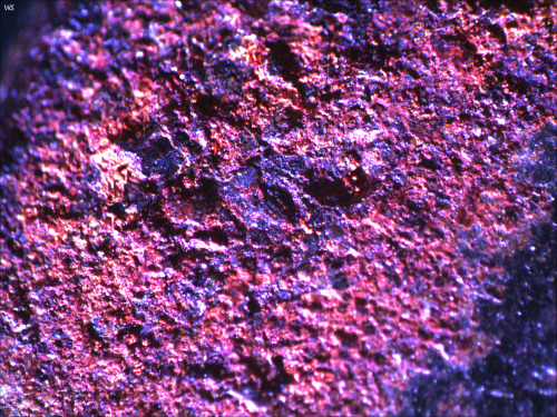
Magnesium oxide single crystals growing on Mg. Courtesy of Anasori Babak, Materials Science and Engineering Dpt, Drexel University, Philadel
Post link
Immunostaining of planktonic Cnidaria. Acetylated tubulin (green), myosin (red), nuclei (blue). Image taken with ZEISS Lightsheet Z.1 during the EMBO course on Marine Animal Models in Evolution & Development, Sweden 2013. www.zeiss.com/lightsheet Sample courtesy of Helena Parra, Institut de Biologia Evolutiva (CSIC-Universitat Pompeu Fabra), Barcelona.
Post link
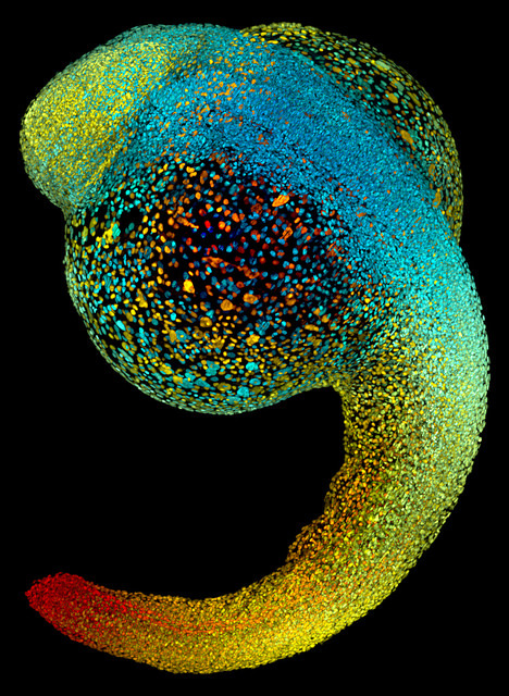
Just 22 hours after fertilization, this zebrafish embryo is already taking shape. By 36 hours, all of the major organs will have started to form. The zebrafish’s rapid growth and see-through embryo make it ideal for scientists studying how organs develop. Image courtesy of Philipp Keller, Bill Lemon, Yinan Wan and Kristin Branson, Janelia Farm Research Campus, Howard Hughes Medical Institute, Ashburn, Va. Part of the exhibit Life:Magnified by ASCB and NIGMS.
Post link
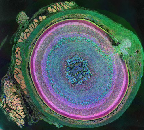
The incredible complexity of a mammalian eye (in this case from a mouse) is captured here. Each color represents a different type of cell. In total, there are nearly 70 different cell types, including the retina’s many rings and the peach-colored muscle cells clustered on the left. Image
Post link
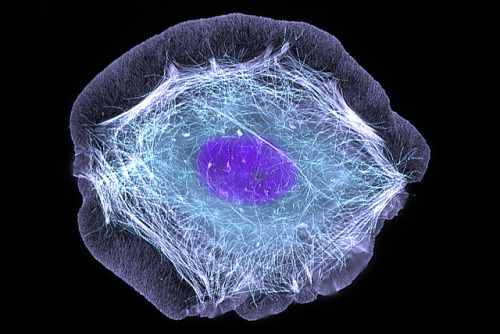
This normal human skin cell was treated with a growth factor that triggered the formation of specialized protein structures that enable the cell to move. We depend on cell movement for such basic functions as wound healing and launching an immune response. Image courtesy of Torsten Wittmann, University of California, San Francisco. Part of the exhibit Life:Magnified by ASCB and NIGMS.
Post link
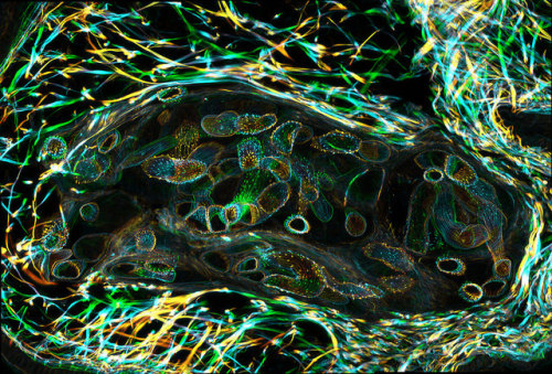
The parasitic worm that causes schistosomiasis hatches in water and grows up in a freshwater snail, as shown here. Once mature, the worm swims back into the water, where it can infect people through skin contact. Initially, an infected person might have a rash, itchy skin or flu-like symptoms, but the real damage is done over time to internal organs. Image courtesy of o Wang and Phillip A. Newmark, University of Illinois at Urbana-Champaign.
Post link
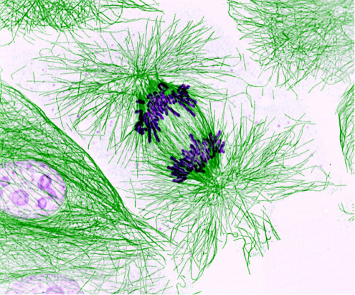
This pig cell is in the process of dividing. The chromosomes (purple) have already replicated and the duplicates are being pulled apart by fibers of the cell skeleton known as microtubules (green). Studies of cell division yield knowledge that is critical to advancing understanding of many human diseases, including cancer and birth defects. Image courtesy of Nasser Rusan, National Heart, Lung, and Blood Institute, National Institutes of Health.
Post link
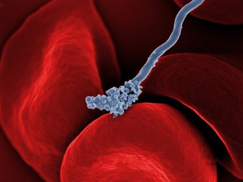
The long, spiral-shaped bacterium (gray) in this image causes relapsing fever, a disease characterized by recurring high fevers, muscle aches and nausea. The relapses result from the bacterium’s unusual ability to change the molecules on its outer surface, allowing it to dodge the human immune system. The disease is transmitted through the bite of a tick (not the same species that transmits Lyme disease) and is found in parts of the Americas, the Mediterranean, central Asia and Africa. Image courtesy of Tom Schwan, Robert Fischer and Anita Mora, National Institute of Allergy and Infectious Diseases, National Institutes of Health.
Post link
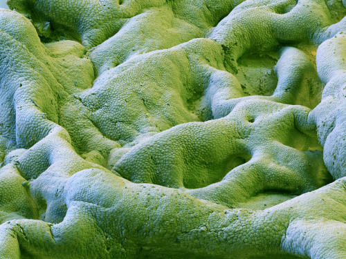
Image SS21025699 (Human Gallbladder SEM)
Scanning electron micrograph (SEM) of the surface of the gallbladder. Each of the small elevations on the wrinkled surface is an epithelial cell.
The gallbladder is a pear-shaped mucosal sac that serves as a reservoir for the bile fluid produced in the liver. Bile is released into the stomach after ingestion, where it plays an important role in the digestion of fats.
The bile emulsifies fats in partly digested food, thereby assisting their absorption. Bile consists primarily of water and bile salts, and also acts as a means of eliminating bilirubin, a product of hemoglobin metabolism, from the body.
Medicines and other “waste products” filtered out by the liver are also contained in the bile and are excreted in this way.
The gallbladder can be affected by gallstones, formed by material that cannot be dissolved, usually cholesterol or bilirubin. These may cause significant pain, particularly in the upper-right corner of the abdomen.
Magnification is 140x at a size of 12x12cm.
Image above © Eye of Science / Science Source
Post link


