#scanning


Discarded AFM tips.
Atomic Force Microscopy, or AFM, is a technique by which a small mechanical probe is scanned across a sample to create a height map. This technique has very high resolution, less than a nanometer, depending on what kind of tip is being used, and can be done in ambient conditions (no need for vacuum). AFM is useful for getting roughness data and measuring film thickness, and can be combined with other microscopy techniques to get a complete picture of your device.
AFM probes often get damaged or dirty, resulting in “tip graveyards” like the one shown here.
Post link
THIRST
Online musical presentation from label Fractal Fantasy for a track by Xzavier Stone is notable for great photorealistic graphics running in a browser. Put together by AlteredQualia with 3D scanning from Chris Rawlinson, it is pushing forward the visual identity for musical releases.
You can try the interactive experience yourself here, as well as view some YouTube uploads of other new tracks by the artist here


3ddd.party
Latest development of digital dancing project from FuzzyWobble now features a portable lightweight 3D scanner for participants to be converted into 3D models, which can be used for party visuals (and even AR):
3DDD.PARTY is a recent experiment involving 3D body scanning & real-time multidimensional visuals.
Multidimensional possibilities include:
→ Real-time VJ Performance ←
→ AR/VR Experiences ←
→ Brand Contextualization ←
→ Hyper Dynamics ←
→ Fictional Textures ←
The system is hireable for events - you can find out more here
Post link
Colour through the Eye of a Mantis Shrimp by ZEISS Microscopy
Via Flickr:
The retina and underlying optic lobes of a mantis shrimp have been stained to reveal the many different units of light information processing before it reaches the central brain. Alexa Fluor probes on fixed mantis shrimp brain section embedded in VectorShield H1000 and imaged with ZEISS LSM 710 confocal microscope www.zeiss.com/lsm Courtesy of Elise Roberts, Honours Student, Marshall Lab, Queensland Brain Institute QBI

Magnesium oxide single crystals growing on Mg. Courtesy of Anasori Babak, Materials Science and Engineering Dpt, Drexel University, Philadel
Post link

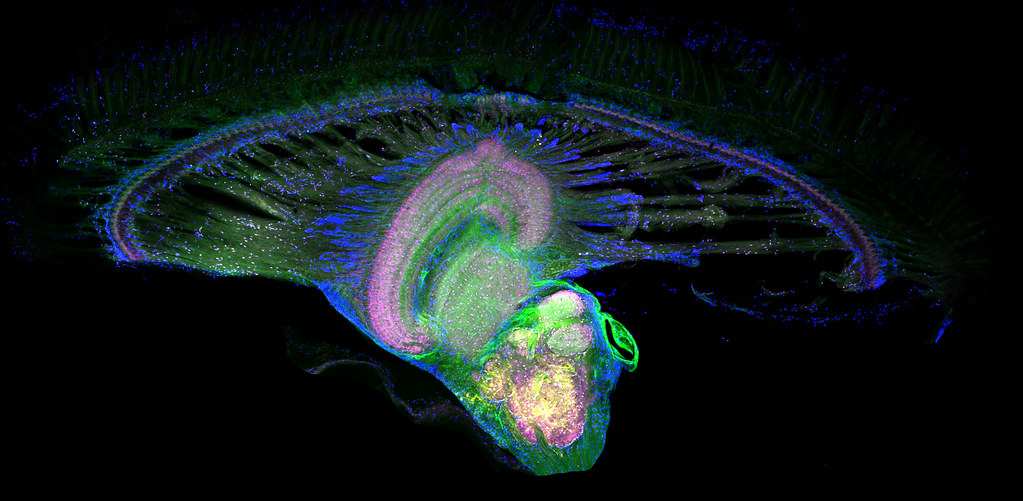
Just 22 hours after fertilization, this zebrafish embryo is already taking shape. By 36 hours, all of the major organs will have started to form. The zebrafish’s rapid growth and see-through embryo make it ideal for scientists studying how organs develop. Image courtesy of Philipp Keller, Bill Lemon, Yinan Wan and Kristin Branson, Janelia Farm Research Campus, Howard Hughes Medical Institute, Ashburn, Va. Part of the exhibit Life:Magnified by ASCB and NIGMS.
Post link

The incredible complexity of a mammalian eye (in this case from a mouse) is captured here. Each color represents a different type of cell. In total, there are nearly 70 different cell types, including the retina’s many rings and the peach-colored muscle cells clustered on the left. Image
Post link
 | ZEISS MicroscopyThis normal human skin cell was treated with a growth fac")
This normal human skin cell was treated with a growth factor that triggered the formation of specialized protein structures that enable the cell to move. We depend on cell movement for such basic functions as wound healing and launching an immune response. Image courtesy of Torsten Wittmann, University of California, San Francisco. Part of the exhibit Life:Magnified by ASCB and NIGMS.
Post link

The parasitic worm that causes schistosomiasis hatches in water and grows up in a freshwater snail, as shown here. Once mature, the worm swims back into the water, where it can infect people through skin contact. Initially, an infected person might have a rash, itchy skin or flu-like symptoms, but the real damage is done over time to internal organs. Image courtesy of o Wang and Phillip A. Newmark, University of Illinois at Urbana-Champaign.
Post link
 and cell skeleton (green) | ZEISS MicroscopyThis pig cel")
This pig cell is in the process of dividing. The chromosomes (purple) have already replicated and the duplicates are being pulled apart by fibers of the cell skeleton known as microtubules (green). Studies of cell division yield knowledge that is critical to advancing understanding of many human diseases, including cancer and birth defects. Image courtesy of Nasser Rusan, National Heart, Lung, and Blood Institute, National Institutes of Health.
Post link

Yeast make bread, beer and wine. And like us, yeast can reproduce sexually. A mother and father cell fuse and create one large cell that contains four offspring. When environmental conditions are favorable, the offspring are released, as shown here.
Post link

Asteroidea Electrica |ZEISS Microscopy
This is a false colored low magnification electron micrograph of free standing graphene foam. Graphene foam is made by growing a few layers of graphene on the surface of a porous metal foam skeleton using chemical vapour deposition technique. The metal foam skeleton is then removed by carefully dissolving it in an etching solution. Because of its unique properties, e.g. electrically conductive, highly porous, and lightweight, graphene foam has the potential to be used in numerous advanced applications including chemical sensing, energy storage, and ultra-lightweight structures. Submitted by Adrianus Indrat Aria.
Post link


About to start another project. I’m standing at the edge, the jump off point. This undertaking feels almost too big, bigger than me. So, I thought it might be a good idea to explore my roots first and find the kernel, the origin space that all of this came from. I’m trying to pull a little nod from the past that it is indeed okay to move forward into an abstract future.
Post link


