#microscope

A water flea.
Female copepod with eggs.
Pond sample.
Quick copepod
Water flea?
Pond sample.
Pond sample. Any guesses on what this organism may be?
Creek sample. Organism eating algae.
Algae and microorganisms from creek sample.
")
24 hours in the life of a zebrafish embryo
In this video you can watch 24 hours (the second day) in the development of a tiny zebrafish embryo.
Some cells have been labelled with a fluorescent protein naturally found in jellyfish. This fluorescent label marks several developing organ systems including the eye, hatching gland and kidney.
You can also see two groups of cells migrating along the trunk of the embryo, depositing smaller clumps of sensory cells as they go. These cells are the precursors of the lateral line system, which gives fish a sense of ‘touch at a distance’, enabling them to shoal, avoid obstacles and find prey.
Look more closely to see individual cells moving around over the skin.
Light sheet microscopy illuminates the specimen with a thin sheet of light and allows scientists like Dr Tanya Whitfield and her team at the University of Sheffield to observe cells and embryos at high speeds or for long times. This research is helping them to gain a better understanding of the amazing process of embryogenesis, including the development of sensory systems such as the lateral line and inner ear.
Video: Sarah Baxendale and Nick van Hateren.
See more beautiful images of zebrafish embryos here


A two-day-old zebrafish embryo brain
These striking images show a zebrafish embryo brain at just two-days-old as seen with a Light Sheet Fluorescent Microscope.
Light sheet microscopy illuminates the specimen with a thin sheet of light and can be used to see relatively large structures in fine detail. You can see all the nerve cells and connections in the zebrafish brain stained with an antibody in red.
Being able to see embryos in this detail allows research teams at the University of Sheffield to follow the amazing process of embryogenesis, from a single cell (the fertilised egg) to a functioning animal with hundreds of different cell types.
Images: Sarah Baxendale, Stone Elworthy and Nick van Hateren.
Post link

Discarded AFM tips.
Atomic Force Microscopy, or AFM, is a technique by which a small mechanical probe is scanned across a sample to create a height map. This technique has very high resolution, less than a nanometer, depending on what kind of tip is being used, and can be done in ambient conditions (no need for vacuum). AFM is useful for getting roughness data and measuring film thickness, and can be combined with other microscopy techniques to get a complete picture of your device.
AFM probes often get damaged or dirty, resulting in “tip graveyards” like the one shown here.
Post link

Bender-scope
The microscopes of the future are all surly and a little bit hungover.
Current mood: “Bite my shiny metal a$$“

“These donuts may eventually have medical applications as active materials.”
Microscopic, 3-D printed donuts, coated in nickel and platinum may be the key to mimicking biological behaviors and delivering drugs or stir samples in labs-on-a-chip — a miniature device that mimics a full laboratory on a microchip.
Read more at Penn State News.
Post link

“Within the in-between” reveals brain cells and their complex interwoven processes. To create this image varying colours have been used to reflect the changing depths of the neuronal processes as they extend through the brain. This image was captured at high-resolution in 3D using state-of-the-art fluorescence microscopy while I was @qldbraininstitute
//
Beneath the Surface opens tonight @po8gallery and includes a series of 5 limited edition large format prints.
100% of the profit from the sale of these prints will go directly towards Schizophrenia research
To find out more and to shop these prints visit the link in my bio ⬆️
.
.
.
.
#neuroscience #microscopy #beautifulscience #sciart #brain #microscope #micro #neuron #beneaththesurface
Post link
.")
.")
.")


.")
.")
Gf: What’s this white stuff on the corals?
Me: no idea
Gf: it looks like what was on my hands yesterday before they swelled up
Me as I bolt excitedly to the other room: I’LL GET THE SLIDES AND THE MICROSCOPE!

SNAILS! Not sure exactly what kind as gf got a free snail from work that no one could identify, but I’m 90% certain it’s some kind of nerite snail. Unfortunately he’s laying eggs on the duncans. Definitely not ideal!


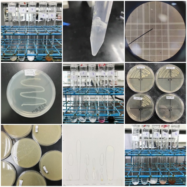
Here are some pictures that I’ve taken from my past laboratory experiments during the whole foundation year + my first year of BSc (Hons) Biotechnology in university! Had some pictures that are from failed / unsuccessful experiments (ㆀ˘・з・˘)
A microscopic timelapse of snowflakes growing