#cardiology

LIVE
I wish I could learn to apreciate more things I have. I remember loving to study medicine, but at th

I wish I could learn to apreciate more things I have. I remember loving to study medicine, but at the same time complaining so much that I couldn’t just enjoy the journey. Now that I have time to rethink everything I hope just to not make the same mistake twice.


Post link

For a while now I have been having palpitations on and off for no particular reason. I started checking my heart rate, using a built-in app on my phone, on a somewhat daily basis shortly after my IBD diagnosis back in 2016 and noticed my resting heart rate was always above what the app said was normal for someone of my age/height/weight/gender. I didn’t think much of it because I was on a lot of meds that, for all I knew, could be causing the increase in heart rate. It was always high whenever I was admitted to the hospital too but I assumed that was due to being in hospital as well as being unwell at the time.

Skip forward to just after my first surgery, and my resting heart rate was always above 120bpm. For reference, for an average adult of my height/weight/age/gender, the resting heart rate is typically between 60 & 80bpm. I figured the 120bpm was a direct result of my surgery and after a few weeks of slowly building my weight back up, my resting heart rate dropped to around 90bpm or so.

From there, I didn’t really notice it much; it became sort of like background noise. I’d notice my heart beating really fast during and after exercise but didn’t really think much of it then either because that’s meant to happen, right?

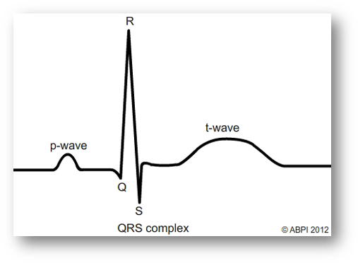
Anyway, after having surgery for the fourth time back in July last year, I started getting sudden onset palpitations with fast breathing and a somewhat panicked feeling (a panic attack?). Again, for no apparent reason. This would last for about 2 minutes before fading, leaving me feeling drained and anxious, so I finally went to my GP. She referred me to have a 24 hour monitor, which involves wearing a heart monitor (similar to an ECG) for 24 hours that records the wearers heart rate which is then analysed by a cardiologist.

After having the 24 hour test at the beginning of the year, I finally saw a cardiologist in clinic earlier this week. They did an ECG and an ultrasound on my heart and, thankfully, everything looks normal. My heart infrastructure is fine and looks healthy, and aside from the palpitations and high resting heart rate, there doesn’t appear to be anything wrong. They don’t want to try any intense treatment unless it starts interfering with my daily life but they did recommend trying beta-blockers to help regulate my heart rate. Because beta blockers can make you tired, the cardiologist said I wouldn’t have to take them all the time but could try taking them whenever I start getting palpitations.

My GP has already filled a prescription for me so I guess I’ll give them a go once they’re ready. Whether this is related to my IBD or not, I don’t know, but it wouldn’t be too much of a stretch to think so as IBD can effect many parts of the body, not just the bowel. I’ll update again on this after I’ve been taking the beta blockers for a bit.

In other news, I’m going to flying to the Netherlands later this week so expect a post about that later next week. I’ll be sure to take note of how going through security goes this time compared to last time (hopefully, it’ll go just as smoothly).

biomedicool:

HEARTBEATS!!

The pause is to allow the atria to fully empty into the ventricle.

Heartbeat on an ECG trace

P Interval (Ventricular Diastole)

  • Atria and ventricles are relaxed
  • blood is flowing into the atria from the veins. 
  • Atrial pressure increases above that of the ventricle, AV valves open allowing blood to flow into the ventricle

P Wave (Atrial Systole) P-Q

Signal transduction from SA to AV nodes. 

  • SA node fires 
  • Atria contract causing atrial systole 
  • which forces all blood into the ventricles
  • emptying the atria.

Q Interval (End of Ventricular Diastole)

Depolarisation of interventricular (IV) septum 

  • AV valves remain open - all remaining blood squeezed into the ventricles. 
  • impulse from the SA node reaches the AV node 
  • whichspreads the signal throughout the walls of the ventricles via bundles of His and Purkinje fibres
  • R peak is the end of ventricular diastole and the start of systole.

R Interval (Ventricular Systole)

Ventricular contraction

  • All blood is now within the ventricles
  • so pressure is higher than in the atria - AV valves close
  • ventricles start to contract although pressure is not yet high enough to open the SL (semilunar) valves

ST Segment (Ventricular Systole)

Ventricular contraction

  • Pressure increases until it equals Aortic pressure,
  • SL valves open
  • blood is ejected into the Aorta (and pulmonary artery) as ventricles contract
  • At this time the atria are in diastole and filling with blood returning from the veins.
  • plateau in ventricular arterial pressure

T Wave (Ventricular Diastole)

T= moment of Ventricular repolarisation immediately before ventricular relaxation

  • Ventricles relax
  • ventricular pressure is once again less than the aortic pressure 
  • so SL valves close

Cardiology Echo Quiz/Case: Which Echo-Sign do you see here and what is y…

Cardiology Echo Quiz/Case: Heart stops beating Part 1! SCD, VT, SVT, AVNRT?

Comment on this video on our Youtube-Channel: https://youtu.be/0BhAcmMroz0

cyberchondria:

Mitral valve prolapse and regurgitation

Nice Loops!

Want more Cardiology Cases & Quizzes?

Visit:https://www.youtube.com/channel/UCJ72o05eM8ZhqTvwf8Gs-SA

Cardiology & Radiology Quiz/Case: Xray+Cardiac Devices. Can you spot them all? Pacemaker, ICD,…?

Watch and comment on Youtube: https://youtu.be/09Kp_wJvvWM

#cardiac    #cardio    #cardiology    #cardiophile    #cardiacdevices    #echocardiography    #pacemaker    #mitraclip    #occluder    #watchman    #amulet    #amplatzer    #cardiologist    #kardiologie    #echokardiographie    #röntgen    #thorax    #radiology    #radiologie    #echocardiogram    
56 y/o F h/o CHF (EF 20-25%), rheumatic heart dz w/ severe mitral stenosis and regurg presents with

56 y/o F h/o CHF (EF 20-25%), rheumatic heart dz w/ severe mitral stenosis and regurg presents with acute onset SOB and light headedness.

Top EKG– before.
Bottom EKG after metoprolol and cardizem.

Diagnosis?


Post link
mynotes4usmle:Cardio Mnemonics for Review Heart Sounds Heart Murmurms  Murmurs & Maneuvers mynotes4usmle:Cardio Mnemonics for Review Heart Sounds Heart Murmurms  Murmurs & Maneuvers mynotes4usmle:Cardio Mnemonics for Review Heart Sounds Heart Murmurms  Murmurs & Maneuvers mynotes4usmle:Cardio Mnemonics for Review Heart Sounds Heart Murmurms  Murmurs & Maneuvers

mynotes4usmle:

Cardio Mnemonics for Review

  1. Heart Sounds
  2. Heart Murmurms 
  3. Murmurs & Maneuvers

Post link

merging and trying to get it “more or less synched”, there must be an easier way to do this..

cardiology
cardiology
loading