#mitosis


instagram.com/p/Bl-7EmXAyFi/
watch this one dog become FOUR DOGS!


Becoming.
byJan van IJken. I want this playing on loop on my gravestone.
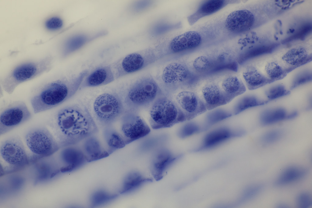
Mitosis - Onion Root Tip, l.s., 100x by Ken Schwarz
Via Flickr:
Amscope PS100-A - #55 Nikon Plan APO 100/1.40 @ 1.05 Achr/Apl oiled Canon 5DMk2


Top: Drawings of mitosis in newt cells found in W. Flemming, Zellsubstanz, kern und zelltheilung (Verlag Vogel, Leipzig, 1882). (A to J) During prophase (A to C) the chromosomes form within the nucleus from a substance termed “chromatin” because of its affinity for dyes. After nuclear envelope breakdown (D), the chromosomes interact with the two separating “centrosomes” (E) to form a spindle-shaped structure (E and F). After the chromosomes attach to the spindle, they become positioned on its equator, halfway between the two poles (G). Once this “metaphase” stage is achieved, the two chromatids comprising each chromosome disjoin and move toward the opposing poles (G and H). During the final stages of mitosis, neighboring chromosomes within the two groups fuse to form the daughter nuclei (H and I), and the cell becomes constricted between them (I) by cytokinesis. (J) Drawing from Schrader’s (2) book depicting conspicuous chromosomal (kinetochore) fibers during early anaphase inLilium.
Bottom: (A to H) Fluorescence micrographs of mitosis in fixed newt lung cells stained with antibodies to reveal the microtubules (green), and with a dye (Hoechst 33342) to reveal the chromosomes (blue). The spindle forms as the separating astral MT arrays, associated with each centrosome (A to C), interact with the chromosomes. Once the chromosomes are segregated into daughter nuclei (F and G), new MT-based structures known as stem-bodies form between the new nuclei (G). These play a role in cytokinesis (H).
Post link

Human epithelial cell in mitosis, fluorescently labeled for alpha tubulin, gamma tubulin and DNA.
Post link


Mi-fourth-sis
Dividing pyrotechnics!
Don’t forget to review the stages of mitosis while watching the fireworks this weekend!
It looks like this one is in metaphase!
Happy 4th of July!


