#hepatic
Kitty Liver
Check meowt!
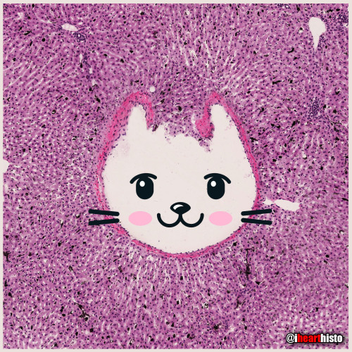
I’m a purrfect hepatic vein surrounded by hepatic lobules!
This is a hematoxylin and eosin stained slice through a liver that was injected with carbon/ink prior to fixation.
The liver is compose of numerous roughly hexagonal shaped lobules. Each lobule has a venule at its center (the central vein; top right) and a series of hepatic triads at its periphery. A triad is a collection of three structures - in this case branches of the hepatic artery, hepatic portal vein and hepatic duct.
Blood in the hepatic artery (oxygenated) and hepatic portal vein (rich in nutrients absorbed from the intestines) travels in sinusoids (the many narrow white spaces in this image) towards the central vein of each lobule. On its way through the sinusoids the blood is processed by the the many hepatocytes that line this region (the pink cells).
Within the sinusoids reside many liver macrophages (Kupffer cells) that phagocytose debris traveling through sinusoids. Normally these cells are invisible in standard H&E preparations but recall that this tissue was injected with ink! The ink was phagocytosed by the macrophages filling their cytoplasm with carbon so that they are now visible as black cells in the sinusoids.
Once the blood enters the central vein of each hepatic lobule they drain into the hepatic veins (kitty!) until the blood reaches the inferior venal cava.
In this way all blood, rich in raw nutrients and toxins absorbed from the GI tract is processed by the liver before it enters the systemic circulation!
Pawsitively unbeliverable!
Post link
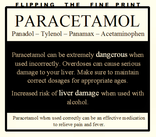
Please stick to correct dosages, I’ve seen too many kids be put on a liver transplant list in the last year - this message is long overdue!
Too many of us think that panadol is harmless and can’t hurt us. It really, really can!
Apologies if there are more common names around the world - I’m in Australia!
Post link
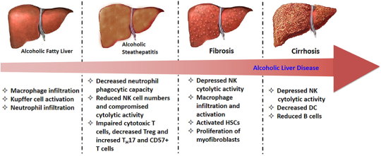
Pathogenesis
Stage One
- Ethanol is metabolised in the liver toacetaldehydeby alcohol dehydrogenase
- which is in turn metabolised to acetate by aldehyde dehydrogenase.
- As acetaldehyde is formed, NAD is metabolised to NADH as a cofactor in this reaction.
- ThisNADH inhibits the actions of isocitrate dehydrogenase and alpha ketoglutarate and fatty acid oxidation, meaning it favours fatty acid synthesis.
- Fat builds up in vacuoles of hepatocytes and mitochondria begin to dysfunction under oxidative stress, creating reactive oxygen species.
- This fatty stage is known as steatosis and is stage one of alcoholic liver disease. It is reversible.
Stage Two
- Inflammatory cytokines such as IL-6 and TNFa influx in, causing inflammation.
- Water begins to accumulate inside hepatocytes and they begin to balloon. This causes significantcholestasis.
- The inflammatory cytokines stimulate hepatocytes to produce collagen, which can begin to formfibrosis.
- This collagen can be pericellular or around central veins, forming collagen bridges. This is a precursor for cirrhosis.
Stage Three
- As the fibrosis progresses,hepatocytes begin to die and cirrhosis occurs.
- This is end-stage alcoholic liver disease and can lead to liver failure and death.

Liver function tests
- Instage 1; all LFTs will be raised. AST and ALT will be raised by approximately 5-10x the upper reference limit except albumin. They will both usually be below 300IU/L. AST will be raised above ALT; this is different to other hepatic conditions such as cholestasis, tumours, hepatitis, where ALT will be above AST.
- In stage 2; ALP and bilirubin will increase significantly as cholestasis occurs and blocks bile duct. This will usually be above 300IU/L, which is different from filtrative diseases, where ALP is usually raised to 200-250IU/L. AST and ALT may increase further.
- Instage 3; AST and ALT are between 10-100x the upper reference limit, suggesting significant cirrhosis and damage to cells. AST will remain above ALT. GGT will be raised significantly, which is common with alcoholic liver disease. Albumin will be decreased, suggestive of total liver failure. In other hepatic conditions, liver damage usually does not reach this stage and so albumin stays within the range.


