#pathologists
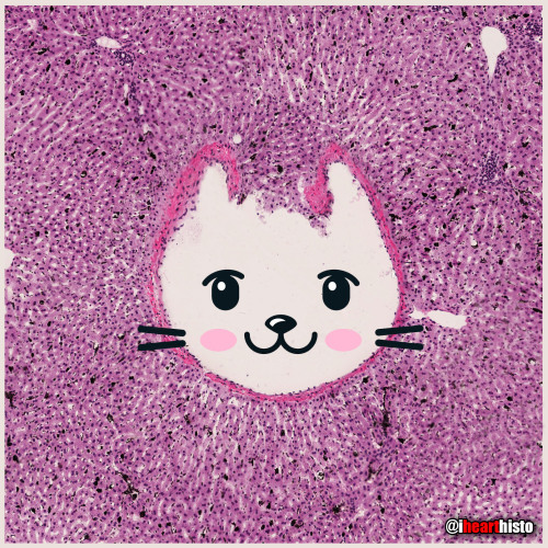
Kitty Liver
Check meowt!
I’m a purrfect hepatic vein surrounded by hepatic lobules!
This is a hematoxylin and eosin stained slice through a liver that was injected with carbon/ink prior to fixation.
The liver is compose of numerous roughly hexagonal shaped lobules. Each lobule has a venule at its center (the central vein; top right) and a series of hepatic triads at its periphery. A triad is a collection of three structures - in this case branches of the hepatic artery, hepatic portal vein and hepatic duct.
Blood in the hepatic artery (oxygenated) and hepatic portal vein (rich in nutrients absorbed from the intestines) travels in sinusoids (the many narrow white spaces in this image) towards the central vein of each lobule. On its way through the sinusoids the blood is processed by the the many hepatocytes that line this region (the pink cells).
Within the sinusoids reside many liver macrophages (Kupffer cells) that phagocytose debris traveling through sinusoids. Normally these cells are invisible in standard H&E preparations but recall that this tissue was injected with ink! The ink was phagocytosed by the macrophages filling their cytoplasm with carbon so that they are now visible as black cells in the sinusoids.
Once the blood enters the central vein of each hepatic lobule they drain into the hepatic veins (kitty!) until the blood reaches the inferior venal cava.
In this way all blood, rich in raw nutrients and toxins absorbed from the GI tract is processed by the liver before it enters the systemic circulation!
Pawsitively unbeliverable!
Post link
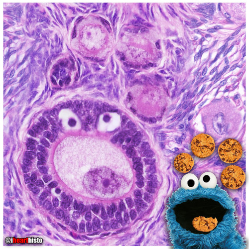
Cookie Monster Ovary
Today’s ihearthisto is brought to you by the letter O for Oocyte!
This is image shows a high magnification view of a slice through an ovary.
You are looking at four tiny primordial follicles (the cookies above Cookie Monster’s head) located in the outer cortex of the ovary. Each of these follicles contains a single dormant, immature egg (a primary oocyte) that is halted in the the first phase of meiosis (prophase I). Eventually these follicles will be recruited to enter the maturation cycle that, against all the odds, could see them develop into an embryo.
Cookie monster himself is a larger multilaminar primary follicle (the cookie in his mouth is the nucleus of the oocyte within the follicle). This type of follicle has already been recruited and it’s follicular cells are dividing and differentiating. The oocyte within it though is still halted in prophase I of meiosis.
An oocyte will only complete meiosis if it is ovulated and fertilized by a spermatozoon (a sperm cell).
Post link

The Cytology Zoo
You can come too, too, too!
An exhibition of smears featuring some familiar looking squamous epithelial cell critters.
Can you recognize them all?
Cytology is the study of individual cells that have been isolated from a specimen. The technique is used mainly to study or screen for cancer but can be useful in the diagnosis of other diseases too, like those involving infectious organisms.
Most of the cells seen in a cytological smear or specimen are obtained in one of three ways:
- Scraping/Brushing the surface of a tissue (this is how cells are obtained from the uterine cervix during a pap smear)
- Collecting a fluid, this could be urine, mucus, blood or semen
- Fine-needle aspirations, this is when cells are removed by sucking them through a fine diameter needle. Fluid from the abdominal, pleural or pericardial cavities are obtained in this way and also cerebrospinal fluid during a lumbar puncture (or spinal tap).
The cells seen here are all squamous epithelial cells that are obtained from the cervix by scraping/brushing.
Credits:
@oreoimc (1-4) & (7-9)
@isac.p (5)
@sza_jhycto (6)
#histology #science #pathology #pathologists #anatomy #autopsy #zoo #animals #cytology #dentalschool #iquizhisto #premed #biology #medicaleducation #meded #nurse #nursing #medschool #medstudent #medicine #medlab #vetscience #vetschool #vetstudent #histologia #histotech #histo #pathArt #ihearthisto

Bender-scope
The microscopes of the future are all surly and a little bit hungover.
Current mood: “Bite my shiny metal a$$“
Jurassic Pork
The pork tapeworm Taenia solium or a brachiosaurus?
i♡histo
This image shows the tapeworm T. solium, a parasite found in the bowels of humans who eat under-cooked pork.
You can see the proglottid segments of the worm’s neck/body and its terrifying scolex (head) complete with a hooked rostellum which it uses to attach itself onto your intestinal wall so that you don’t poop it out easily.
Fully grown worms can expand to around 2-3 meters in length and are composed of numerous segments each of which form their own reproductive unit.
The life cycle of the parasite begins when pigs ingest the T. solium eggs (from fecal contamination). The eggs develop into larvae inside the pig & then migrate through the intestinal wall to form cysts in their muscles & tissues.
Once slaughtered for meat, the larvae can be ingested by humans if the pork is eaten raw or under-cooked. Once in the human small intestine the larvae grow into large adult worms where they persist often unnoticed due to the lack of symptoms associated with their existence. The worms lay eggs into the intestines which are released during defecation thus continuing the life cycle.
“Life finds a way” - Dr. Ian Malcolm, Jurassic Park
A major complication of infection with T. solium is Cysticercosis. This is a parasitic tissue infection caused by the larval cysts of the tapeworm. These larval cysts infect brain, muscle, or other tissue, and are a cause of adult onset seizures. A person only gets cysticercosis by swallowing the eggs found in the feces of a person who has an intestinal tapeworm NOT from contracting a tapeworm by eating under-cooked pork itself. So people living in the same household with someone who has a tapeworm have a much higher risk of getting cysticercosis than people who don’t because they can be exposed to the eggs released in the stool.
Post link
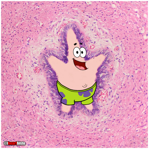
⭐ Patrick Star in a Sperm Tube
My favorite Patrick in honor of #stpaddysday☘️
Some say that Patrick Star and the male reproductive tract are fairly similar… but if you look closely there’s actually a vas deferens.
This image shows the tube through which spermatozoa travel during ejaculation. It is called the vas deferens (ductus deferens).
Histologically it is unique because of its unique pseudostratified epithelium (surrounding Patrick…check out the unique regular beaded appearance of the basal cells in this epithelium) and its triple-layered smooth muscle wall - so thick you can actually palpate it within the spermatic cord within the scrotum. Patrick is actually within the empty space of the lumen of the tube (where the sperm travels).
Spermatozoa are produced in the seminiferous tubules of the testes and travel to the epididymis where they complete the maturation process. During ejaculation, a peristaltic wave of strong muscular contractions propel the sperm from the epididymis and into the vas deferens. This wave of peristalsis continues along the triple-thick muscle wall of the vas deferens eventually pushing the sperm out into the urethra. From here they have enough momentum to travel along the length of the penis before exiting exit the tube at the external urethral meatus (the fancy anatomical name for the hole at the end of the penis).
But sperm are not the only stuff that is ejaculated… at the same time as the sperm is moving along this pathway, the smooth muscle surrounding a number of glands also contracts which pushes their secretions into the urethra too at the same moment that the sperm is moving through it. These include secretions from the seminal vesicles (seminal fluid), prostate gland (prostatic fluid) and bulbourethral glands.
The final concoction that is ejaculated is actually a combination of sperm and all of these extra sperm-nourishing secretions and collectively it is known as semen.
Which brings us back to where we started with this sea-man - Patrick Star!
#histology #science #pathology #pathologists #anatomy #autopsy #patrickstar #spongebob #vasdeferens #malereproductive #stpatricksday #menshealth #premed #biology #medicaleducation #meded #nurse #nursing #medschool #medstudent #medicine #medlab #vetscience #vetschool #ihearthisto
Post link


