#microscopy
Check out the June issue of SciArt Magazine @sciartglobal for a feature on my recent work with @po8gallery and @artisanqld + work by @kelly_heaton @GDunnArt @NickErvinck and others. Link in their bio
•
White gold ring from Beneath the Surface with Australian parti sapphire and pink sapphire accent
•
#neuroscience #microscopy #sciart #sciencejewelry #contemporaryjewellery #australiandesigner #organicjewelry #madebyhand #australiansapphire
Post link
Light pink morganite set into an 18ct white gold band and surrounded by a 18ct and 14ct yellow gold fans. Part of Beneath the Surface, the rings in this series reflect both underwater forms and the fusion of synaptic vesicles with the cell surface in neurons. View this piece @po8gallery
•
#neuroscience #microscopy #beautifulscience #sciart #sciencejewelry #contemporaryjewellery #australiandesigner #organicjewelry #madebyhand #australiansapphire
If you’re in Melbourne today for #marchforscience stop by @po8gallery and see Beneath the Surface. The exhibition includes large format images from within the brain created @qldbraininstitute and Cerulean Odyssey, a large silver object created to raise money and awareness for motor neurone disease research (MND/ALS).
∙
My colleagues and other researchers in Australia work long hours in positions that often have poor long term job security to push forward our understanding of ourselves, our planet, and create cures and treatments for diseases that you may one day find yourself afflicted with. It is essential but under funded and under recognised work. We all benefit from the discoveries made by the generations of scientists who lived before us, we should celebrate the scientists carrying on this tradition
∙
We are also raising money for schizophrenia research through sale of limited edition prints linked to the exhibition - link in bio ☝️
∙
#neuroscience #microscopy #beautifulscience #sciart #sciencejewelry #contemporaryjewellery #australiandesigner #organicjewelry #madebyhand #mnd #als #schizophrenia
Post link
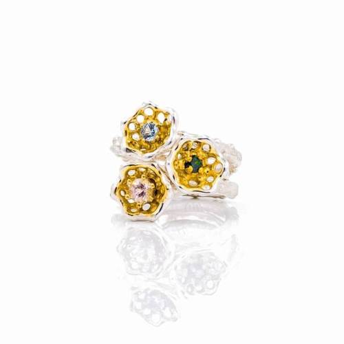
Cusp rings, to be worn alone or stacked together. Open silver frameworks cup around bright aquamarine, Australian sapphire and light pink morganite
Part of Beneath the Surface, exhibiting with @po8gallery
#neuroscience #microscopy #beneaththesurface #sciart #sciencejewelry #contemporaryjewellery #australiandesigner #organicjewelry #madebyhand #australiansapphire
Post link
My colleagues and I have just published a new paper in Molecular Neurobiology which contributes important insights into how vitamin D deficiency during embryonic development can alter the brain’s dopamine system.
•
This research is just a part of the important work coming out of the Queensland Brain Institute linking vitamin D to autism traits and schizophrenia. Working with Dr. Leon Luan and Prof. Darryl Eyles was one of the highlights of my time @qldbraininstitute and I’m looking forward to seeing the rest of this work being published soon.
•
This image is one of test images I captured when developing the new analysis methods for these studies. We combine advanced microscopy and analysis techniques to image large areas of the brain and characterise each individual brain cell
•
#neuroscience #microscopy #microscopic #beautifulscience #imaging #microphotography #micrography #brain #scienceisart #sciart
Post link
Thank you to everyone who came and supported the opening of Beneath the Surface!
Creating this collection took countless hours at the bench in solitude so it was great enjoying your beautiful city, making new friends and seeing some familiar faces. Grateful to be able to share my work with you thanks to the amazing team @po8gallery ✌️
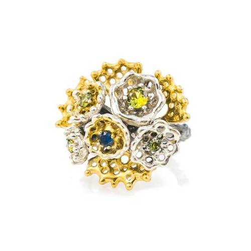
// This is one of the emerging rings featuring Australian sapphires, exhibition open until the 29th
.
.
.
#neuroscience #microscopy #beautifulscience #sciart #sciencejewelry #contemporaryjewellery #australiandesigner #organicjewelry #madebyhand #australiansapphire
Post link
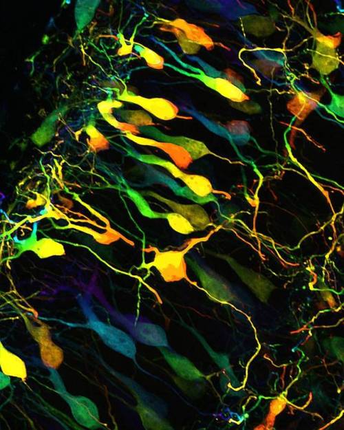
“Within the in-between” reveals brain cells and their complex interwoven processes. To create this image varying colours have been used to reflect the changing depths of the neuronal processes as they extend through the brain. This image was captured at high-resolution in 3D using state-of-the-art fluorescence microscopy while I was @qldbraininstitute
//
Beneath the Surface opens tonight @po8gallery and includes a series of 5 limited edition large format prints.
100% of the profit from the sale of these prints will go directly towards Schizophrenia research
To find out more and to shop these prints visit the link in my bio ⬆️
.
.
.
.
#neuroscience #microscopy #beautifulscience #sciart #brain #microscope #micro #neuron #beneaththesurface
Post link
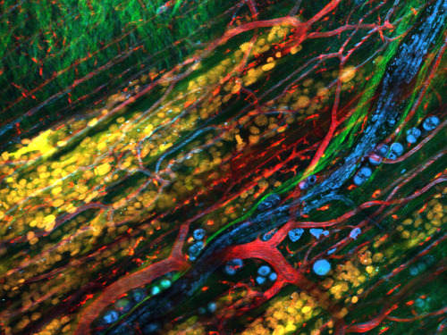
Collective invasion of B16/F10 melanoma cells into mouse dermis, detected by infrared multiphoton microscopy. Tumor cells expressing E2-Crimson and Histone-2B-GFP are (false-colored) yellow (cytoplasm) and blue (nuclei). Nerve fibers and fat cells are cyan (third harmonic) and collagen bundles and muscle fibers are red (second harmonic), respectively. Blood vessels and phagocytic cells are labeled with TM-Rhodamine-dextran in green. By courtesy of Bettina Weigelin and Peter Friedl, NCMLS, Radboud University, Nijimegen, Netherlands.
(Source:https://www.lavisionbiotec.com/applications/oncology.html)
Alexander, S., Weigelin, B., Winkler, F., Friedl, P., Preclinical intravital microscopy of the tumour-stroma interface: invasion, metastasis, and therapy response, Curr Opin Cell Biol. (2013), 25(5), 659-71
Post link
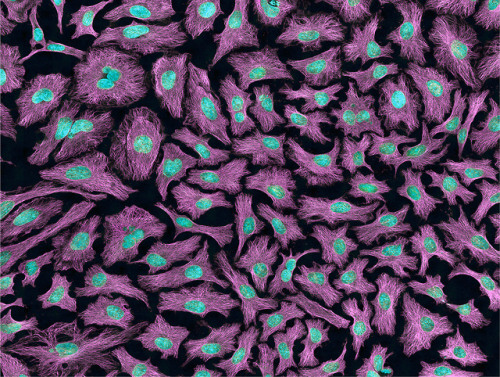
Multiphoton fluorescence image of HeLa cells with cytoskeletal microtubules (magenta) and DNA (cyan). Nikon RTS2000MP custom laser scanning microscope.
Source: National Center for Microscopy and Imaging Research (NCMIR) (source)
Credit Line: National Center for Microscopy and Imaging Research
Post link
Fig. 4. Overexpression of LGP85 does not cause enlargement of lysosome, but results in formation and accumulation of the late endosome-lysosome hybrid organelle. To label lysosomes, COS cells were incubated with Texas-Red dextran for 4 hours and chased for 20 hours. After that, cells were transiently transfected with LGP85 and fixed at 12 (A-C), 24 (D-F), and 36 (G-I) hours after transfection, followed by staining for LGP85 (A,D,G, green). Cells were visualized by confocal microscopy. The right columns show the merged images of LGP85 (green) and dextran (red). Bars, 20 μm.
A role for the lysosomal membrane protein LGP85 in the biogenesis and maintenance of endosomal and lysosomal morphology
Toshio Kuronita, Eeva-Liisa Eskelinen, Hideaki Fujita, Paul Saftig, Masaru Himeno, Yoshitaka Tanaka
Journal of Cell Science 2002 115: 4117-4131; doi: 10.1242/jcs.00075
Post link
#Lipilight#MemBright and super resolution #LiveSR on living muscle cells! by Bruno Cadot
Visit website : https://cadotbruno.com
Visit Bruno’s Twitter : https://twitter.com/cadotbru
Post link
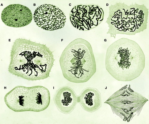
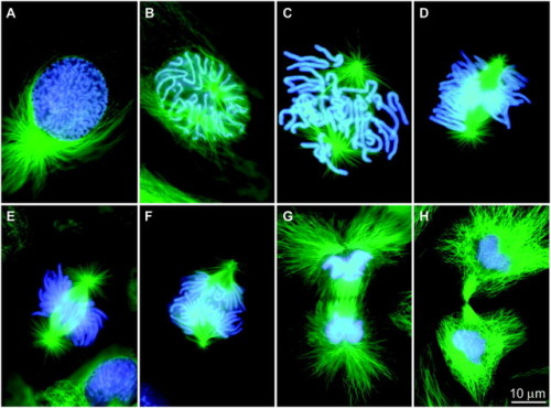
Top: Drawings of mitosis in newt cells found in W. Flemming, Zellsubstanz, kern und zelltheilung (Verlag Vogel, Leipzig, 1882). (A to J) During prophase (A to C) the chromosomes form within the nucleus from a substance termed “chromatin” because of its affinity for dyes. After nuclear envelope breakdown (D), the chromosomes interact with the two separating “centrosomes” (E) to form a spindle-shaped structure (E and F). After the chromosomes attach to the spindle, they become positioned on its equator, halfway between the two poles (G). Once this “metaphase” stage is achieved, the two chromatids comprising each chromosome disjoin and move toward the opposing poles (G and H). During the final stages of mitosis, neighboring chromosomes within the two groups fuse to form the daughter nuclei (H and I), and the cell becomes constricted between them (I) by cytokinesis. (J) Drawing from Schrader’s (2) book depicting conspicuous chromosomal (kinetochore) fibers during early anaphase inLilium.
Bottom: (A to H) Fluorescence micrographs of mitosis in fixed newt lung cells stained with antibodies to reveal the microtubules (green), and with a dye (Hoechst 33342) to reveal the chromosomes (blue). The spindle forms as the separating astral MT arrays, associated with each centrosome (A to C), interact with the chromosomes. Once the chromosomes are segregated into daughter nuclei (F and G), new MT-based structures known as stem-bodies form between the new nuclei (G). These play a role in cytokinesis (H).
Post link
Widefield fluorescence microscopy of Paramecium spec. before (left) and after (right) deconvolution.
Source : Leica Microsystems
Post link
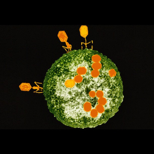
Coloured transmission electron micrograph (TEM) of T2 bacteriophage viruses (orange) attacking an Escherichia coli bacterium. Each phage consists of a large DNA- containing head and a tail composed of a central sheath with several fibres. The fibres attach to the host cell surface, and the phage DNA is injected into the cell through the sheath. It instructs the host to build copies of the phage (progeny, in cell). Destroying the host’s DNA releases nucleotide building blocks, from which phage DNA is synthesised. It is then packaged in the progeny, which are released when the host bursts. Magnification: x64,000.
Post link
Stem cells in the skin - stem cells labelled in red are found in special microenvironments where they are surrounded by other types of cell.
Post link
A rendered image of a primary neuronal stem cell culture in which cells were labeled with different fluorescently labeled proteins that differentiate between stem cells (orange/yellow) and their neuronal ‘offspring’ (blue/ green/ purple).
Post link
Huntington’s stem cell derived oligodendrocyte precursors stained for phalloidin (green), vinculin (red) and DNA (blue).
Post link
Mouse diaphragm consisting of neurons, muscle cells and neuromuscular junctions under the confocal microscope: Green structures show neurons (Alexa 488), red areas are neuromuscular junctions (rhodamin), and blues areas are muscle fiber (myosin, DODT contrast).
Post link
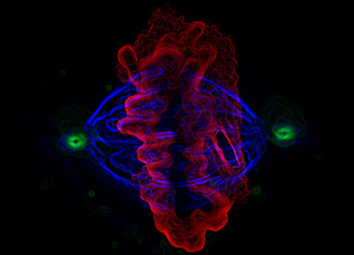
Human epithelial cell in mitosis, fluorescently labeled for alpha tubulin, gamma tubulin and DNA.
Post link
Nearly 2K followers on tumblr! wtf is happening? ;) and now something for this occasion:
Photomicrograph of a midsaggital section showing the different components of the rat cerebellum, including Purkinje neurons in green, glia (non-neuronal cells) in red, and cell nuclei in blue. This image is part of an ongoing effort to develop methods to enable the creation large-scale digital atlases of the brain.
Post link
Magnesium oxide single crystals growing on Mg. Courtesy of Anasori Babak, Materials Science and Engineering Dpt, Drexel University, Philadel
Post link


