#microscopic
Water flea moving about
Cladocera (water fleas) and air bubbles.
Cladocera, a small crustacean.
A water flea.
Female copepod
Normally invisible, these dynamic branches and tubulations are a just a few of the many internal compartments of a single cell. They’ve been made to glow by using green fluorescent protein found in jellyfish
.
When I was studying neuroscience all I wanted to do was get involved in research on consciousness, but I took a side step and started an imaging project in a cancer biology lab with Prof. Jenny Stow @uniofqld This is when I first started using microscopes to image fluorescent proteins in cells.
Its one thing to know that without your awareness every single cell in your body is almost vibrating with activity and something entirely different to actually see it with your own eyes. Looking down into an ocean of darkness and seeing these glowing worlds alive within cells was a profound experience that completely captured my imagination. A few years later I started work establishing an imaging facility @qldbraininstitute so these techniques could be used to study the brain. It’s been such an incredible journey to see this technique move from allowing us to see inside a single cell, like in this movie I captured over a decade ago, to watching every neuron in a brain flashing with activity.
.
This dynamic intracellular activity and the even smaller structures that surround these tubules and spheres helped inspire the jewellery and objects I’m exhibiting with @po8gallery this month. Opening night Thursday 6th. Come and see!!
.
.
#neuroscience #microscopy #beautifulscience #sciart #microscopic #fluorescence #contemporaryjewellery #australiandesigner #organicjewelry #madebyhand
My colleagues and I have just published a new paper in Molecular Neurobiology which contributes important insights into how vitamin D deficiency during embryonic development can alter the brain’s dopamine system.
•
This research is just a part of the important work coming out of the Queensland Brain Institute linking vitamin D to autism traits and schizophrenia. Working with Dr. Leon Luan and Prof. Darryl Eyles was one of the highlights of my time @qldbraininstitute and I’m looking forward to seeing the rest of this work being published soon.
•
This image is one of test images I captured when developing the new analysis methods for these studies. We combine advanced microscopy and analysis techniques to image large areas of the brain and characterise each individual brain cell
•
#neuroscience #microscopy #microscopic #beautifulscience #imaging #microphotography #micrography #brain #scienceisart #sciart
Post link
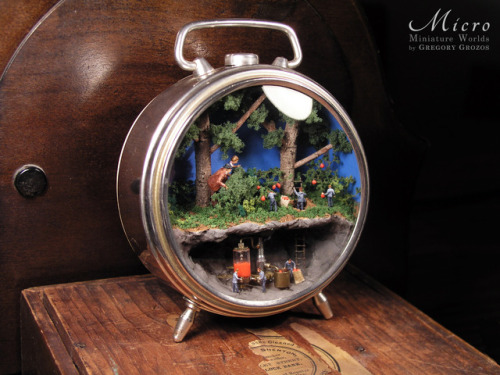
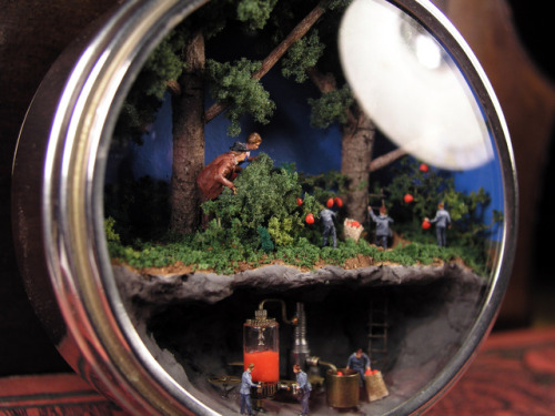
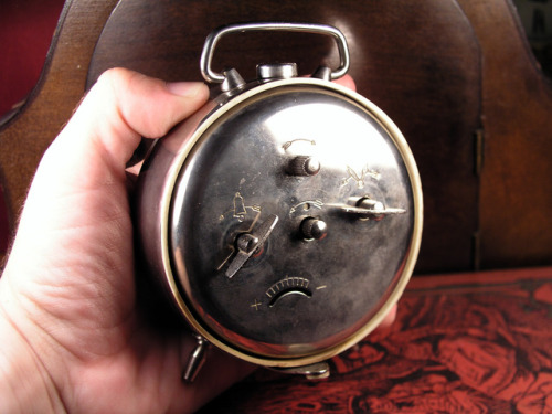
Here’s my latest miniature world, this time built inside in a vintage clock case. A man and a woman stumble upon some tiny little people, who are coming out of a hole in the ground and are gathering the fruits from the surrounding bushes. They then go down the ladder to an underground cavern and feed the fruits inside a machine which turns them into juice, filling up a glass tank. Some of the little people are filling their cups and are tasting the juice.
View more of my work by visiting my Facebook page or Etsy shop:
www.facebook.com/microjewellery
www.etsy.com/shop/MicroJewellery
Post link
This is one of my favorite designs, an entire miniature scene closed in a glass dome ring. The scene includes a town made out of tiny tiny houses, mountains, a lake, the sea, a boat and other microscopic details.
As always you can find available at my Etsy shop:
http://www.etsy.com/shop/MicroJewellery
Post link
Image SS2519384 (Cricket Sound Comb, SEM)
Scanning Electron Microscope image of the sound producing comb of the Field Cricket (Gryllus pennsylvanicus).
This specimen was collected in the Finger Lake Region of New York State. The sound from this insect comes from their comb rubbing against the underside of the opposite wing. Only male crickets produce the characteristic sound.
View More Cricket Sound Comb Images
Some cricket species have several types of chirping songs in their repertoire. The calling song attracts females and repels other males, and is fairly loud.
The courting song is used when a female cricket is near and encourages her to mate with the caller.
A triumphal song is produced for a brief period after a successful mating, and may reinforce the mating bond to encourage the female to lay some eggs rather than find another male.
An aggressive song is triggered by contact chemoreceptors on the antennae that detect the presence of another male cricket.
Image above © Ted Kinsman / Science Source
Post link
Image SS2599851 (Colored SEM Of An Eardrum)
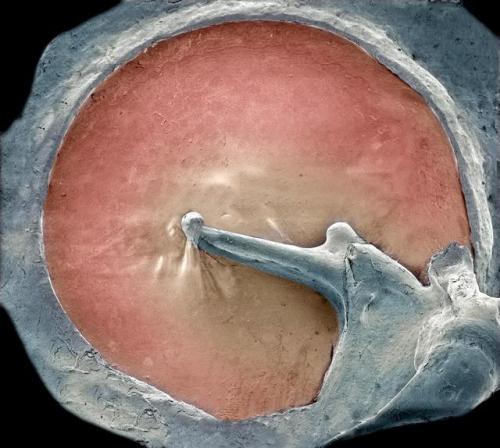
Colored Scanning Electron Micrograph (SEM) of an Eardrum (red).
The eardrum, or tympanic membrane, is located in the middle ear. It is a thin, cone-shaped membrane that separates the external ear from the middle ear
The eardrum transmits sound to the inner ear through three tiny bones called the ossicles (malleus, incus and stapes).
View More Amazing SEMs Of The Inner Ear
The malleus (center) is attached to the inner eardrum. It articulates with the incus (right), which, in turn, links to the stapes (not seen).
The middle ear transforms sound waves into vibrations in the fluid-filled cochlea of the inner ear, which are then sent as nerve signals to the brain.
Magnification: x16 when printed 10 centimeters wide.
© Steve Gschmeissner / Science Source
Post link
Image SS2385580 (Blood Clot, SEM)
Colored scanning electron micrograph (SEM) of a human blood clot.
Here it has formed inside a blood vessel following an injury. Red blood cells (a.k.a. erythrocytes) are seen trapped by a web of brown fibrin threads. The fibrin is an insoluble protein and these threads are an essential mechanism for the arrest of bleeding.
Platelets (a.k.a. thrombocytes) are a component of blood whose function (along with the coagulation factors) is to react to bleeding from blood vessel injury by clumping, thereby initiating a blood clot.
As the blood cells become trapped they lose their normal rounded shape (as at upper left). Blood clots form to repair blood vessels damaged by injury or disease.
Abnormal clotting in intact arteries and veins (as occurs in a thrombus) is the principal cause of heart attack and stroke.
On the other side of the coin, there is a bleeding disorder called, Hemophilia, where the blood does not clot properly. This can lead to spontaneous bleeding as well as bleeding following injuries or surgery.
Magnification: x2500 at 6x6cm size.
©Eye of Science / Science Source
Post link
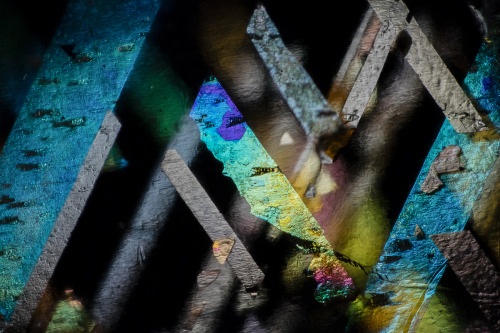
This material is truly special.
Ilmenite and hematite in orthoclase feldspar from Harts Range, Australia.
Field of view = 3mm.
Post link
Here is the watercolor version of the colored pencil drawing i made last week! Im not entirely sure if this is a princess with very intricate insect inspired costuming, or an insect that has captured a human, has her mind-controlled, and is using her as a puppet/continuous source of nutrition (yummy organs)
.
.
.
#art #drawing #illustration #gouache #colorstudy #watercolor #painting #colorpencil #coloredpencil #creepy #insect #microscopic #chromatic #prismacolor #windsornewton #pelikan #bug #parasite #naked #woman #figure #figuredrawing #nakedwoman #princess #queen
Post link
Moldavite, a glowing green Tektite from Czech Republic
Click Here to See More Crystals
Click Here to Shop Crystals
Post link


“No two snowflakes are alike”
Snowflakes caught on black velvet then captured by light sensitive plates
“Under the microscope, I found that snowflakes were miracles of beauty; and it seemed a shame that this beauty should not be seen and appreciated by others. Every crystal was a masterpiece of design and no one design was ever repeated., When a snowflake melted, that design was forever lost. Just that much beauty was gone, without leaving any record behind.”
“A careful study of this internal structure not only reveals new and far greater elegance of form than the simple outlines exhibit, but by means of these wonderfully delicate and exquisite figures much may be learned of the history of each crystal, and the changes through which it has passed in its journey through cloud-land. Was ever life history written in more dainty hieroglyphics!”
Wilson Bentley (1865-1931)
Biography from the Museum of Everything
Wilson Snowflake Bentley was a farmer, entrepreneur, meteorologist and artist, who dedicated his life to the photography of snow.
Born in 1865 to a poor country family in Vermont, Bentley’s imagination was first sparked by his mother’s gift of a microscope on his 15th birthday. Bentley taught himself how to use the contraption, combined it with a Bellow’s camera and embarked upon his creative journey.
By the age of 20, Bentley had taught himself how to immortalise the structure of frozen water on glass plates. Over the next 40 years, he would assemble a vast encyclopaedia of scientific abstraction, photographing several thousands of individual snowflakes and frosts.
Bentley’s achievements led to a greater understanding of the uniqueness, importance and beauty of this natural form. Yet Bentley was far more than a passionate hobbyist. He was a self-styled outlier and adventurer, who realised the potential of microphotography to capture the impossible individuality of each and every crystalline flake.
In his lifetime, Bentley proselytised his discoveries across the United States, sending prints and plates to Museums, Universities and more. The public and press championed his achievements; yet the scientific and cultural communities of the time were often reluctant to welcome the innovations of this pioneering image maker.
Today, over one hundred years later, Bentley’s photographs are celebrated not simply as science, but as art. His microphotographs are featured in public and private collections worldwide, and are regularly curated in exhibitions such as Massimiliano Gioni’s The Keeper at the New Museum in New York (2016) and The Museum of Everything in Rotterdam (2016) and Australia (2017/18).
Wilson Snowflake Bentley died of pneumonia after being caught in a snowstorm in 1931.
Sources:
Snowflake Man Bio — Snowflake Bentley
https://href.li/?https://www.gallevery.com/artists/wilson-bentley#
Here’s Where the Saying ‘No Two Snowflakes Are Alike’ Originated
1982 PHOTOMICROGRAPHY COMPETITION
Lars Bech
Academisch Medisch Centrum
Amsterdam, The Netherlands
Subject Matter:
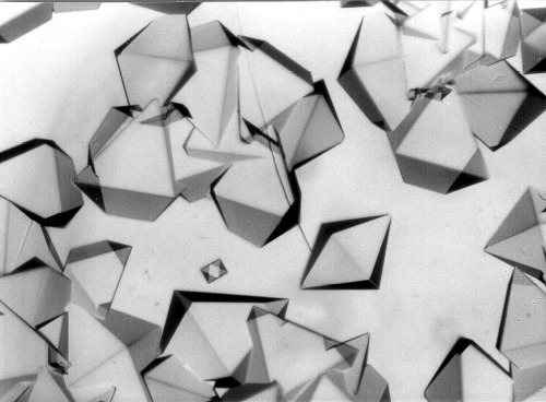
Combination of urea, phosphor acid, ammonia melted
(25x)Technique:
Post link
1993 PHOTOMICROGRAPHY COMPETITION
Douglas Moore
University of Wisconsin - Stevens Point
Stevens Point, Wisconsin, USA
Subject Matter:
Draparnaldia glomerata algae
(200x)Technique:
Post link
A close-up of this leopard moth (Bracca rotundata) shows of its softer side.
Post link
Mineral under pressure
This is a photograph of mineral called pyroxene and was taken with a petrographic microscope, and it captures megascale forces of the tectonic processes on the microscale level. This mineral is about 1.5 mm and was found in a rock called greenschist.
This rock is heavily deformed, sheared, due to the force of the tectonic plates pushing against each other. Imagine one plate being a bread and other a knife, and the rock you are looking at is like a butter being smeared on the toast.
The pyroxene is harder than the rest of the minerals in this rock, and so, as the pressure is applied, the other minerals are deformed and squashed against the pyroxene while it remains largely undamaged.
Example from the Nubra Valley, Ladakh, Himalaya
Post link


