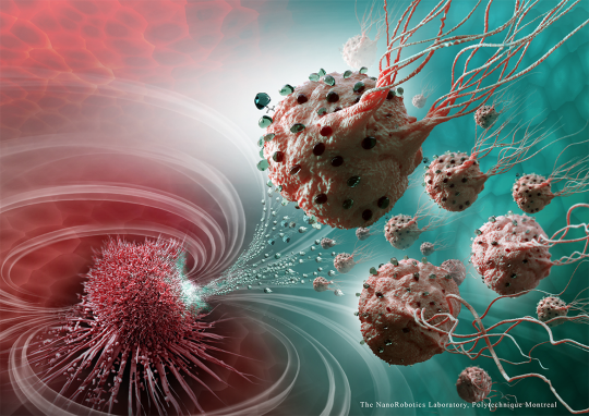
In a major breakthrough for cancer research a team from Polytechnique Montréal, Université de Montréal and McGill University, Canada, have developed nanorobotic agents able to administer drugs with precision to targeted cancerous cells of tumours. Injecting medication in this way ensures the optimal targeting and avoids jeopardising organs and the surrounding tissue.
The nanorobotic agents are made of over 100 million flagellated bacteria, giving them the ability to self-propel. The agents are filled with drugs and move alone the most direct path between the injection point and the area of the body to cure. The propelling force of the agents is enough to travel and enter the tumours.
Once inside a tumour, the agents can detect oxygen-depleted areas, known as hypoxic zones, and deliver the drug to them. Hypoxic zones are resistant to most therapies, including radiotherapy.
The bacteria that make up the agents rely on two natural systems to move around. The synthesis of a chain of magnetic nanoparticles acts as a compass, allowing the bacteria to move in the direction of a magnetic field, while a sensor measuring oxygen concentration enables them to reach active tumour regions. The bacteria were exposed to a computer-controlled magnetic field, showing the researchers that it can perfectly replicate artificial nanorobots.
The researchers say that the nanorobots open the door for the synthesis of new vehicles for therapeutic, imaging and diagnostic agents, as well as having use in chemotherapy by eliminating the harmful side effects by targeting the affected area.
Berkeley Lab scientists show how tiny, metal-rich particles can be excited with a low-power laser for deep-tissue imaging
A research team has demonstrated how light-emitting nanoparticles, developed at the U.S. Department of Energy’s Lawrence Berkeley National Laboratory (Berkeley Lab), can be used to see deep in living tissue.
The specially designed nanoparticles can be excited by ultralow-power laser light at near-infrared wavelengths considered safe for the human body. They absorb this light and then emit visible light that can be measured by standard imaging equipment.
The development and biological imaging application of these nanoparticles is detailed in a study published online Aug. 6 in Nature Communications.
Researchers hope to further develop these so-called alloyed upconverting nanoparticles, or aUCNPs, so that they can attach to specific components of cells to serve in an advanced imaging system to light up even single cancer cells, for example. Such a system may ultimately guide high-precision surgeries and radiation treatments, and help to erase even very tiny traces of cancer.
Hundreds of polymers that could kill drug-resistant superbugs in novel ways can be produced and tested with light, using a method developed at the University of Warwick. The new methodology may identify antimicrobials for a range of applications from personal care to coatings.
Researchers from the Department of Chemistry and Warwick Medical School developed a way to synthesise large libraries of polymers to make screening for antimicrobial activity faster, and without the need to use sealed vials.
By using multiple ‘building blocks’ in their polymers, new antimicrobials were identified – some of which appear to inhibit bacteria growth, contrary to predictions. The benefit of the method is that it allows screening of hundreds of different structures, enabling the researchers to 'go fishing’ for new properties, which in this case was antibiotic activity.
Antimicrobials are essential not just in the treatment of internal disease and infections, but also in personal care products, such as contact lenses or shampoo, in foods, or as topical creams.
These tiny interwoven fibers make up the 3-D fabric “scaffold” into which a strong, pliable hydrogel is integrated and infiltrated with stem cells, forming a framework for growingcartilage. The resulting composite material is tough, flexible and formable and has excellent frictional properties. It mimics both the strength and suppleness of native cartilage.
Visit Website | Image credit: Frank Moutos, Orthopaedic Research Laboratories, Duke University, and Farshid Guilak, Professor of Orthopaedic Surgery and Mechanical Engineering and Materials Science, Duke University
A wearable device that monitors compounds in your sweat for up to a week could help in the early detection of diabetes, according to the University of Texas, USA, research team.
The wearable device, pictured above, can detect cortisol, glucose and interleukin-6 – interconnected compounds linked to diabetes – in perspired sweat. ‘If a person has chronic stress, their cortisol levels increase, and their resulting insulin resistance will gradually drive their glucose levels out of the normal range. At that point, one could become pre-diabetic, which can progress to type 2 diabetes,’ said Dr Shalini Prasad, Professor of Bioengineering.
Not only is the Texas team’s device functional for one week without loss of signal integrity, it requires a far smaller degree of sweat – one to three microlitres, rather than 25 to 50 – to be effective. Prasad said, ‘We spent three years producing that evidence. At those low volumes, the biomolecules expressed are meaningful. We can do these three measurements in a continuous manner with that little sweat.’
Bioprinting is an additive manufacturing process similar to 3D printing – it uses a digital file as a blueprint to print an object layer by layer. But unlike 3D printing, bioprinters print with cells and biomaterials, creating organ-like structures that let living cells multiply.