Heated magnetic nanoparticles may be the future of eradicating cancer cells without harming healthy tissue, according to research from the University of Buffalo, USA. The nanoparticles strike tumours with significant heat under a low magnetic field.
Hao Zeng, Professor of Physics at Buffalo, said, ‘The main accomplishment of our work is the greatly enhanced heating performance of nanoparticles under low-field conditions suitable for clinical applications. The best heating power we obtained is close to the theoretical limit, greatly surpassing some of the best performing particles that other research teams have produced.’
Targeting technologies would first direct nanoparticles to tumours within the patient’s body. Exposure to an alternating magnetic field would prompt the particles’ magnetic orientation to flip back and forth hundreds of thousands of times a second, causing them to warm up as they absorb energy from the electromagnetic field and convert it to thermal energy.
Two particles have been tested – manganese-cobalt-ferrite and zinc ferrite. While the manganese particle reached maximum heating power under high magnetic fields, the biocompatible zinc ferrite was efficieny under an ultra-low field.
While this form of treatment, known as magnetic nanoparticle hyperthermia, is not new, the Buffalo-designed particles are able to generate heat several times faster than the current standard.
The vials shown here contain a molecule that researchers can activate with light to affect biological processes in mammals. It’s part of a class of potential smart drugs that are under development to treat ailments including diabetes, blindness, and the side effects of chemotherapy. Because the molecules turn on only when hit with a certain wavelength of light, researchers can control when and where the compounds are active in the body. This photo was taken by Dusan Kolarski, a graduate student at the University of Groningen in the lab of Ben Feringa, who won the 2016 Nobel Prize in Chemistry for his work on molecular machines. Feringa’s group is working to create antibiotics that can be activated with light and thus may pose a lower risk of bacterial resistance. The team also wants to make water-soluble motors for applications in photopharmacology.—ALEXANDRA TAYLOR
In the 17th Century two giants of science, Isaac Newton and Robert Hooke, were both trying to understand how the wings of butterflies and peacocks, which are made of the same material as our fingernails and hair, could colors of such brilliant quality. They both came to the same conclusion, the color was a result of tiny structures on the wing, structures so small that they could not observe it themselves but had deduced must exist.
Science and technology have progressed far in those 300 years and not only can we easily observe the structure of a butterfly’s wing that produces such brilliant color, but we can readily create them ourselves. Inspired by this kind of structural color, researchers at Kyoto University’s Institute for Integrated Cell-Material Science (iCeMS), led by Prof. Easan Sivaniah, in collaboration with researchers from Semmelweis University and Kyoto University Medical School, have produced a structural color device for measuring the beating of heart cells which they hope will help speed up the process of pharmaceutical testing.
Like the wing of a butterfly, this device produces structural color from micro-patterns developed on the surface of a polymer gel. Heart cells beating on the device cause the structural color to change which can be detected easily with low power microscopes.
The complex architecture of bone is challenging to recreate in the lab. Therefore, advances in bone tissue engineering (BTE) aim to build patient-specific grafts that assist bone repair and trigger specific cell-signaling pathways. Materials scientists in regenerative medicine and BTE progressively develop new materials for active biological repair at a site of defect post-implantation to accelerate healing through bone biomimicry.
Rapidly initiation of new bone formation at the site of implantation is a highly desirable feature in BTE, and scientists are focused on fabricating grafts that strengthen the material-bone interface after implantation. Bioactive glass can bond with bone minutes after grafting, and silk fibroin, a natural fibrous protein has potential to induce bone regeneration. Hybrid materials that exploit these properties can combine the osteogenic potential and the load-bearing capacity for potential applications in large-load bone defect models.
In a recent study, Swati Midha and co-workers developed a novel 3-D hybrid construct using silk-based inks with different bioactive glass compositions integrated to recreate a bone-mimetic microenvironment that supports osteogenic differentiation of bone marrow mesenchymal stem cell (BMSC) lines in the lab. Now published in Biomedical Materials, IOP Science, the scientists used direct writing instruments to produce the silk fibroin-gelatin-bioactive glass scaffolds (SF-G-BG). The results delivered appropriate cues to regulate the development of customized 3-D human bone constructs in vitro.
A team of researchers led by Biomedical Engineering Professor Sam Sia at Columbia Engineering has developed a way to manufacture microscale-sized machines from biomaterials that can safely be implanted in the body. Working with hydrogels, which are biocompatible materials that engineers have been studying for decades, Sia has invented a new technique that stacks the soft material in layers to make devices that have three-dimensional, freely moving parts. The study, published online January 4, 2017, in Science Robotics, demonstrates a fast manufacturing method Sia calls “implantable microelectromechanical systems” (iMEMS).
By exploiting the unique mechanical properties of hydrogels, the researchers developed a “locking mechanism” for precise actuation and movement of freely moving parts, which can provide functions such as valves, manifolds, rotors, pumps, and drug delivery. They were able to tune the biomaterials within a wide range of mechanical and diffusive properties and to control them after implantation without a sustained power supply such as a toxic battery. They then tested the “payload” delivery in a bone cancer model and found that the triggering of release of doxorubicin from the device over 10 days showed high treatment efficacy and low toxicity, at 1/10 of the standard systemic chemotherapy dose.
A skin-like polymeric material is using carbon nanotubes (CNTs) to bring a sense of touch to robotic and prosthetic devices. Developed by researchers at Stanford University and Xerox Palo Alto Research Center, the flexible, polymeric skin or ‘digital tactile system’ (DiTact) incorporates CNT pressure sensors and flexible organic printed circuits to mimic human response [Tee et al., Science 350 (2015) 313].
‘‘We wanted to make a sensor skin that communicates in the same way as the body,’’ explains research student Alex Chortos, one of the lead authors of the work. ‘‘The goal is to make skin for prosthetics that can feel touch in a natural way and communicate that information to the person wearing the prosthetic device.’’
In the body, receptors in the skin relay sensing information directly to the brain in a series of voltage pulses rather like Morse code. Artificial devices employ tactile sensing to improve the control of neuroprosthetics and relieve phantom limb pain. But, to date, prosthetic skin devices have had to use a computer or microprocessor to turn the output from sensors into a signal compatible with neurons.
The new approach, by contrast, combines these operations in a single system of piezoresistive pressure sensors embedded in a flexible circuit layer. The sensors are made from a CNT composite dispersed in a flexible polyurethane plastic and molded into pyramidal structures. The pyramidal shape is crucial because it allows the pressure range of the sensor to be tuned to that of skin.
Self-assembling nanosphere clusters may improve everything from drug synthesis to drug delivery
Miniature self-assembling “flasks” created at the Weizmann Institute may prove a useful tool in research and industry. The nanoflasks, which have a span of several nanometers, or millionths of a millimeter, can accelerate chemical reactions for research. In the future, they might facilitate the manufacture of various industrial materials and perhaps even serve as vehicles for drug delivery.
Dr. Rafal Klajn of the Weizmann Institute’s Organic Chemistry Department and his team were originally studying the light-induced self-assembly of nanoparticles. They were employing a method earlier developed by Klajn in which inorganic nanoparticles are coated in a single layer of organic molecules that change their configuration when exposed to light; these alter the properties of the nanoparticles such that they self-assemble into crystalline clusters. When spherical nanoparticles of gold or other materials self-assembled into a cluster, empty spaces formed between them, like those between oranges packed in a case. Klajn and his team members realized that the empty spaces sometimes trapped water molecules, which led them to suggest that they could also trap “guest” molecules of other materials and function as tiny flasks for chemical reactions. A cluster of a million nanoparticles would contain a million such nanoflasks.
As reported in Nature Nanotechnology, when the scientists trapped molecules that tend to react with one another inside the nanoflasks, they found that the chemical reaction ran a hundred times faster than the same reaction taking place in solution. Being confined inside the nanoflasks greatly increased the concentration of the molecules and organized them in a way that caused them to react more readily. Enzymes speed up chemical reactions in a similar manner – by confining the reacting molecules within a pocket.
For all the good they do, eye drops and ointments have one major drawback: It’s hard to tell how much of the medication is actually getting to the eye. Now in a study appearing in ACS Applied Materials & Interfaces, scientists report that they have developed a contact lens that changes color as drugs are released. This visual indicator could help eye doctors and patients readily determine whether these medications are where they should be.
Eyes are adept at keeping things out. When something ventures into or toward an eye, the lids blink and tears start rapidly flowing to avoid infection and damage from foreign objects. These processes are usually helpful, but they can hinder the uptake of much-needed medications. Studies suggest that less than 5 percent of drugs in eye drops and ointments are absorbed, and much of the absorbed medication ends up in the bloodstream instead of the eye, causing side effects. Contact lenses may be a more effective way to deliver drugs directly to the eye, but real-time monitoring of drug release is still a challenge. So Dawei Deng and Zhouying Xie sought to create a drug-delivering contact lens that would change color as the medication is released into the eye.
Techniques could lead to personalized wearable and implantable devices
Hearing aids, dental crowns, and limb prosthetics are some of the medical devices that can now be digitally designed and customized for individual patients, thanks to 3-D printing. However, these devices are typically designed to replace or support bones and other rigid parts of the body, and are often printed from solid, relatively inflexible material.
Now MIT engineers have designed pliable, 3-D-printed mesh materials whose flexibility and toughness they can tune to emulate and support softer tissues such as muscles and tendons. They can tailor the intricate structures in each mesh, and they envision the tough yet stretchy fabric-like material being used as personalized, wearable supports, including ankle or knee braces, and even implantable devices, such as hernia meshes, that better match to a person’s body.
As a demonstration, the team printed a flexible mesh for use in an ankle brace. They tailored the mesh’s structure to prevent the ankle from turning inward – a common cause of injury – while allowing the joint to move freely in other directions. The researchers also fabricated a knee brace design that could conform to the knee even as it bends. And, they produced a glove with a 3-D-printed mesh sewn into its top surface, which conforms to a wearer’s knuckles, providing resistance against involuntary clenching that can occur following a stroke.
“This work is new in that it focuses on the mechanical properties and geometries required to support soft tissues,” says Sebastian Pattinson, who conducted the research as a postdoc at MIT.
Cuts, scrapes, blisters, burns, splinters, and punctures — there are a number of ways our skin can be broken. Most treatments for skin wounds involve simply covering them with a barrier (usually an adhesive gauze bandage) to keep them moist, limit pain, and reduce exposure to infectious microbes, but they do not actively assist in the healing process.
More sophisticated wound dressings that can monitor aspects of healing such as pH and temperature and deliver therapies to a wound site have been developed in recent years, but they are complex to manufacture, expensive, and difficult to customize, limiting their potential for widespread use.
Now, a new, scalable approach to speeding up wound healing has been developed based on heat-responsive hydrogels that are mechanically active, stretchy, tough, highly adhesive, and antimicrobial: active adhesive dressings (AADs). Created by researchers at the Wyss Institute for Biologically Inspired Engineering at Harvard University, the Harvard John A. Paulson School for Engineering and Applied Sciences (SEAS), and McGill University, AADs can close wounds significantly faster than other methods and prevent bacterial growth without the need for any additional apparatus or stimuli. The research is reported in Science Advances.
A new mathematical model describes how highly concentrated antibody solutions separate into different phases, similar to an oil and water mixture. This separation can reduce the stability and shelf-life of some drugs that use monoclonal antibodies, including some used to treat autoimmune diseases and cancer. A team of scientists from Penn State and MedImmune, LLC (now AstraZeneca) investigated the thermodynamics and kinetics, the relationships between temperature, energy, and the rates of chemical reactions, of the phenomenon using an innovative method that allows for the rapid study of multiple samples at once. A paper describing their model appears July 22, 2019, in the journal Proceedings of the National Academy of Sciences.
Many drugs today are stored as solids and dissolved in IV bags for delivery to patients, but the pharmaceutical industry has been moving toward drugs that can be stored as liquids and given via a shot. Some of these drug solutions, like those used to treat autoimmune diseases and some cancers, contain high concentrations of monoclonal antibodies—proteins that attach to foreign substances in the body, like bacteria and viruses, flagging them for destruction by the patient’s immune system.
Advances in 3-D printing have led to new ways to make bone and some other relatively simple body parts that can be implanted in patients. But finding an ideal bio-ink has stalled progress toward printing more complex tissues with versatile functions – tissues that can be loaded with pharmaceuticals, for example. Now scientists, reporting in the journalACS Biomaterials Science & Engineering, have developed a silk-based ink that could open up new possibilities toward that goal.
Most inks currently being developed for 3-D printing are made of thermoplastics, silicones, collagen and gelatin or alginate. But there are limits to how these inks can be used. For example, the temperatures, pH changes and crosslinking methods that may be required to toughen some of these materials can damage cells or other biological components that researchers would want to add to the inks.
Additives, such as cytokines and antibiotics, are useful for directing stem cell functions and controlling infections, respectively. To address these bio-ink limitations, David L. Kaplan and colleagues turned to silk protein and developed a way to avoid these harsh processing conditions.
The researchers combined silk proteins, which are biocompatible, and glycerol, a non-toxic sugar alcohol commonly found in food and pharmaceutical products. The resulting ink was clear, flexible, stable in water, and didn’t require any processing methods, such as high temperatures, that would limit its versatility. The researchers say the novel material could potentially be used in biomedical implants and tissue engineering.
Medical devices designed to reside in the stomach have a variety of applications, including prolonged drug delivery, electronic monitoring and weight-loss intervention. However, these devices, often created with nondegradable elastic polymers, bear an inherent risk of intestinal obstruction as a result of accidental fracture or migration. As such, they are usually designed to remain in the stomach for a limited time.
Now, researchers at MIT’s Koch Institute for Integrative Cancer Research and Massachusetts General Hospital (MGH) have created a polymer gel that overcomes this safety concern and could allow for the development of long-acting devices that reside in the stomach, including orally delivered capsules that can release drugs over a number of days, weeks or, potentially, months following a single administration.
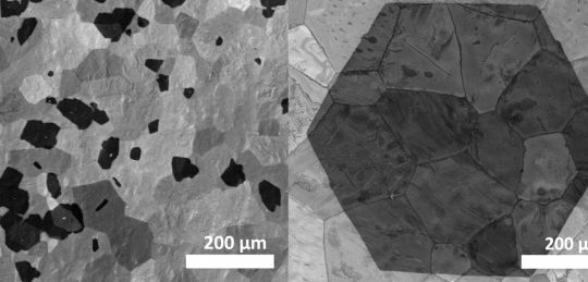
Researchers from the Nanomaterials by Design Group at University of Oxford, led by professor Nicole Grobert have produced millimetre-sized crystals of high-quality graphene in minutes, using a chemical vapour deposition technique (CVD).
The new method produces 2–3mm graphene crystals in 15 minutes, compared to current process which can take up to 19 hours.
Researchers took a thin film of silica deposited on a platinum foil which when heated, reacts to create a layer of platinum silicide. This layer melts at a lower temperature than platinum and silica to create a thin liquid layer that smooth’s out nanoscale ‘valleys’ in the platinum, so that carbon atoms in methane gas brushing the surface form large flakes of graphene.
Grobery, said, ‘Not only can we make millimetre-sized graphene flakes in minutes but this graphene is of a comparable quality to any other methods.’
The team believe the CVD technique could also have additional benefits claiming with a thicker liquid layer to insulate it the graphene might not have to be removed from the substrate before it can be used – an expensive and time consuming process.
Grobert added, ‘Of course a great deal more work is required before we get graphene technology, but we’re now on the cusp of seeing this material make the leap from the laboratory to a manufacturing setting, and we’re keen to work with industrial partners to make this happen.
The researchers hope to develop this technique further and produce flakes of graphene in large wafer-sized sheets.
To find out more on materials science, packaging and engineering news, visit our website IOM3 or follow us on Twitter @MaterialsWorld for regular news updates. You can also now get access to our content any time, anywhere via our app. For more information, visit app.materialsworld.org.
The new synthetic polymer material creates an instant scaffold, sort of like stacked gumballs, that allows new tissue to latch on and grow within the cavities formed between linked spheres of gel.
Conventionally, ointments and other hydrogel dressings have been used to fill in wounds to keep the areas moist and accelerate healing. But none of the materials used now provide a scaffold to allow new tissue to grow while the dressing itself degrades. As a result, the new tissue growth is relatively slow and fragile.
So bringing about an injectable biomaterial that promotes rapid regeneration of tissue has been a “holy grail” in the field of tissue engineering, said co-principal investigator Dino Di Carlo.
They envision the material being useful for a wide variety of wound application, including lacerations to large-area burns.
UC Berkeley researchers have also been developing new approaches to tissue engineering. Last March, their advancement in “herding cells” marked a new direction for smart bandages.
Graphene Ink 3-D Printed Medical Implant Grows Nerve Cells
There is no shortage of excitement for the possibilities of 3-D printing. The manufacturing technique uses a machine that squirts layer upon layer of material to build three-dimensional objects. The prevailing vision for 3-D printing is that one day we’ll be able to make smartphones, sensors, drones or other complex machines right in our homes.
But if we’re ever to have desktop devices that can output things like consumer electronics or novel biomedical devices, there are a number of obstacles that need to be overcome. Today’s consumer units most commonly use hot plastic that quickly solidifies to build shapes. This material is neither particularly strong nor is it electrically conductive, a characteristic necessary to build electronic components into devices.
Researchers all around the world are looking for materials that can unlock some of 3-D printing’s bigger promises. Now Northwestern University researchers say they have created a 3-D printing ink that is stronger, electrically conductive and biocompatible using another material that has been generating much excitement over the last decade–graphene. See more gifs and learn more below.
For the first time, patented titanium fiber plates developed by Japanese engineers for medical use have been tested in an animal model. Researchers from Shinshu University found that, unlike conventional plates, titanium fiber plates do not cause bone embrittlement after close contact with the bone for prolonged periods. This could eliminate the need for plate extraction and the associated surgical risks.
“Ourtitanium fiber plates, unlike conventional titanium plates, are prepared by compressing titanium fibers at normal room temperature into plates without changing the fiber shape,” said Takashi Takizawa, M.D., the paper’s first author from the department of orthopaedic surgery at the Shinshu University School of Medicine. “They can compensate for the major drawback of conventional titanium plates, and find application in a range of fixation and bone tissue repair uses at various sites of the body.”
Their results were published in the January 25th online issue of the journal Advanced Materials.
Most commonly used to hold bones in place while they heal, titanium plates are erosion resistant and strong enough to hold the mending bones in place. Doctors may elect to implant a titanium plate in a patient with a bad fracture, a severe skull injury, or a degenerative bone disease.
Researchers at IIT-Istituto Italiano di Tecnologia fabricated an artificial device reproducing a 1:1 scale model of the blood-brain barrier (BBB), the anatomical and functional structure that protects the central nervous system from external substances, such as contaminants, but also drugs when they are injected intravenously into the body. The device, which is a combination of artificial and biological components, will be fundamental for studying new therapeutic strategies to overcome blood-brain barrier and treat brain diseases, such as tumors.
The study was coordinated by Gianni Ciofani, researcher at IIT in Pontedera (Pisa) and Professor at Politecnico di Torino, in the framework of the research project SLaMM funded by the European Research Council (ERC) and aiming at developing new nanotechnologies for the treatment of brain diseases.
The device is described in a paper published today by the scientific journal Small and highlighted by the journal inside cover: it is a microfluidic device that combines artificial components made with 3-D advanced microfabrication techniques (two-photon lithography) and biological ones, that is endothelial cells (the cells covering blood vessels).
For the first time, researchers have fabricated sensing elements known as fiber Bragg gratings inside optical fibers designed to dissolve completely inside the body. The bioresorbable fiber Bragg gratings could be used for in-body monitoring of bone fracture healing and for safer exploration of sensitive organs such as the brain.
A fiber Bragg grating is an optical element inscribed in an optical fiber, which is widely used as a sensing instrument. Although fiber Bragg gratings are commonly used for applications such as real-time monitoring of the structural health of bridges or tracking the integrity of airplane wings, until now they didn’t exhibit characteristics preferred for use in the body. With a design that allows them to break down similarly to dissolvable stitches, the new glass fibers should be safe for patients even if they accidently break, according to the researchers.
“Our work paves the way toward optical fiber sensors that can be safely inserted into the human body,” said Maria Konstantaki, a member of the research team from the Institute of Electronic Structure and Laser (IESL) of the Foundation of Research and Technology - Hellas (FORTH), Greece, that fabricated and characterized the new gratings. “Because they dissolve, these sensors don’t need to be removed after use and would enable new ways to perform efficient treatments and diagnoses in the body.”
At the recent International Conference on Robotics and Automation, MIT researchers presented a printable origami robot that folds itself up from a flat sheet of plastic when heated and measures about a centimeter from front to back. The MIT researchers’ centimeter-long origami…