#sciblr


An appreciation post for one of my favorite birds, the Anhinga! These birds are good divers and will completely submerge to catch their prey. Since they have no oils on their wings to repel water, anhingas have to stretch out their wings to let them dry before they can fly!
Pictures taken at Huntington Beach State Park, Murrels Inlet, SC
every movie villain scientist: begin human trials
me: what a joke, you have no data. you fraud, no journal will publish you. you aren’t ready for human trials. you are a joke on the scientific method

Additionally:If you’re making a slide-based presentation, consider giving your slides to a non-expert without context. If they can put together your main points and overall narrative from slides alone, then your slides are doing something right!
(From my twitter, which you can find here)I wanted to ask what do you mean by non-expert? Like someone who’s never studied your topic intensely or are we going all the way to someone not in your field or somewhere in between?
Because for most presentations, I feel like someone outside the field entirely not really understanding it is expected. They won’t have the necessary basic vocab (sometimes I babble on about groups or partitions or literally whatever when I’m around non-math friends without expecting them to have any clue what I’m talking about and indeed they don’t). On the other hand, someone else in an adjacent topic or someone a bit younger does sound like a good choice. A couple weeks ago, @counter-example and I practiced conference presentations with each other a couple times which was extra helpful bc we tend to do pretty different math but we both have decent upper level undergrad math backgrounds.
That’s awesome, @sevenfactorial! I’m glad you found someone who provides you useful feedback!
Re: your opening question, I don’t think there’s a universally applicable answer. When I wrote “non-experts”, I had in mind pretty much anyone who hasn’t worked in the subfield of the presentation’s content, but you’re right that there’s likely some minimum overlap of knowledge necessary for useful feedback (a minimum which I imagine relates to the intended audience of the specific presentation; slides for a subfield-specific conference can be more technical than slides for the general public, for example). You’ve pointed out that choosing people in the same field but in a different subfield is a good strategy, and I agree.
I most recently got feedback on my physics slides from a friend who is pursuing a math Ph.D., and his comments were extremely useful despite himself being unfamiliar with a large majority of the physics & jargon. Asking a variety of people seems most useful to me (the more data, the merrier), but as for what single individual would provide the most valuable input? I’m not sure.
I also think it’s important to dial expectations to fit each feedback provider. For example, while my math friend doesn’t know a lot of the vocab that my audience would (so that a critique like “I don’t know what this word means” wouldn’t necessarily warrant action), he picked up that certain things were important from context and thus–even though the details escape him–he could identify the overall narrative. His lack of knowledge about my work is actually a boon: he can keep in mind the bigger picture without getting lost in the details that I might be distracted by. I figure that if he can follow my narrative without understanding the details, then I can be confident my organization will be crystal clear to my audience too. If he mentions that something seems important despitemy intentions, then I can think about reducing my inadvertent overemphasis of that thing.
So I’ll refine my earlier statement to be: I recommend acquiring feedback from multiple people at varying levels of familiarity with your content, while also being careful to gauge that feedback through both the perspective of each feedback provider as well as your eventual intended audience. (I was careful to use plural wording in the original tweet, but dropped it in my text addition on Tumblr, which I would change in retrospect.)
Post link


Annnnd just like that, I’m back from PHENO! Y’all, I had a fantastictime.
I got to talk shop with plenty of physicists (familiar and new), attend excellent presentations, and introduce myself to important people in the field.
Highlight of the Conference: On Tuesday, I presented my current research project. My talk was the second in a coordinated pair of presentations, the first being my advisor’s. His talk allowed me to focus on details and he did a great job of hyping me up along the way, which gave me STRENGTH and ENERGY. I then proceeded to CRUSH my presentation. The question session went well too (people had so many great questions that there wasn’t enough time to answer them all!), and after the talk I received lots of compliments regarding both the project and my delivery. So yeah, it went about as well as possible! Which is made all the better by the fact that several big shots (who’ve done related work) were in the audience too!!
If you’re interested in checking it out, the abstract and slides for my PHENO2019 presentation, titled
** Cancellations in Spin-2 KK Mode Scattering Amplitudes at High Energies **
are online at
** I’m finally in the right frame of mind to polish up this presentation **
I’m grateful to my past self: they pushed against the latest bout of depression + dissociation in order to get it 80% of the way there. And now that the fog is lifting, I can more clearly see an end product in the existing draft.
Today, I complete this task! Let’s get this!!
All right: I’ve drafted next week’s presentation and sent it to my group for feedback. I already know several big changes I want to make, but I’ve also already spent too much time & energy on it, and I promised my group I’d send it yesterday, sooo… yeah. Sometimes ya just gotta call it good and hit send.
The biggest hold-up is that I’m not really feeling my current intro & outro. We’ll see what my group says, I guess? One silver lining is that many of these slides & images are easily ported into future talks, including my eventual thesis defense. By keeping my long-term goals in mind and working hard now, I can streamline future work.
I’ve already profited from that philosophy when it comes to making images. Last year, I converted all of my technical illustration machinery into Photoshop. It was a huge pain at the time, but having everything in Photoshop now made it wayyy easier to edit existing images for this talk.
But, yeah, I can tell I’m burning out on this project, so I need to step back and recharge. I think I’ll write up some generic QFT notes and relax.
I hope you’re doing well, my friend! I’m wishing you the best!
—
Edit, the following day: It turns out the biggest hold-up was actually being in a depressive low. WHOOPS.

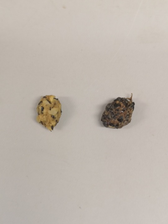
Very cute baby robin. Cat caught and bought into us. The second photo is of two pellets he has produced - the black one on the right from his last meal with his parents - insect shells mostly. The one on the left is from his meals with us - a special mix for growing young passerines. I thought it was very cool to see the diet difference in pellet form!


—December 23rd 2020, 1:54 am.
just a few more days until christmas! i hope everyone’s feeling the holiday spirit despite being stuck indoors. ❣︎
this was my desk setup in studying for my first term finals and i’m so relieved it’s finally over, i think i did pretty good and that makes me happy! i especially love having photos of my friends and family hung up for motivation. ☺︎︎♡︎
i still don’t have a fixed theme for my posts, hehe sorry about that.


—November 21st 2020, 12:30 pm.
hi! i’m kind of back :))
i haven’t been posting in a while and now i’m almost through with the first term of my accelerated med course! there’s definitely been a lot of difficult times but we just got to get through it right? here’s some of my photos that summed up my past few months hehe.
also, meet my doggo mocha! ♡︎


—June 21st 2020, 10:49 am.
i don’t particularly like this spread, to be honest, i just wanted to take pictures of the creamy cinnamon buns before i ate them HAHA. also, i found that mini canvas & easel lying around and i’m hoping to paint something on it soon! any suggestions?
+ my course’s orientation is today! freaked out is an understatement. i’m literally shaking.
Week 13of2020 quarantine challenge
sunday: Where would you rather be from?
this is the easiest question yet — Japan or Korea definitely! i’m just so in love with their weather, people, culture and everything about the place. i visited Japan twice now and Korea once, and both times were just an unforgettable experience! nevertheless, i am happy to be from the ph ♡
but i also wouldn’t mind being in new zealand right now tbh.
Tonsillitis is inflammation of the pharyngeal tonsils.
- Inflammationusually extends to the adenoid and the lingual tonsils
- Most commonly viral
- Most cases of bacterial tonsillitis are caused by group A beta-hemolytic Streptococcus pyogenes (GABHS) - strep throat.
- Spread through the air.
Symptoms may include sore throat, fever, enlargement of the tonsils, trouble swallowing, and large lymph nodes around the neck. Complications include peritonsillar abscess.

Recurrent tonsillitis
- A polymicrobial flora consisting of both aerobic and anaerobic bacteria
- Other competing bacteria are reduced - less interference to GABHS infection.
- Streptococcus pneumoniae, Staphylococcus aureus, and Haemophilus influenzae are the most common bacteria isolated in recurrent tonsillitis, and Bacteroides fragilis is the most common anaerobic bacterium isolated in recurrent tonsillitis.
- The microbiologies of recurrent tonsillitis in children and adults are different; adults show more bacterial isolates, with a higher recovery rate of Prevotella species, Porphyromonas species, and B fragilis organisms , whereas children show more GABHS. Also, adults more often have bacteria that produce beta-lactamase.
Chronic tonsillitis
- Polymicrobial bacterial population present
- There is likely a relationship between tonsillar size and chronic bacterial tonsillitis based on both the aerobic bacterial load and the absolute number of B and T lymphocytes.
- Fewer dendritic cells on the surface epithelium and more in the crypts and extrafollicular areas during chronic tonsillitis.
- Radiation exposure may relate to the development of chronic tonsillitis. A high prevalence of chronic tonsillitis was noted following the Chernobyl nuclear reactor accident in the former Soviet Union.
The tonsils are a set of lymphoid organs consisting of the adenoid tonsil, two tubal tonsils, two palatine tonsils, and the lingual tonsils. These organs play an important role in the immune system.

Palatine tonsil
- Main tonsils
- Non-keratinized stratified squamous epithelium
- Incompletely encapsulated
- Long, branched crypts
- Located on sides of oropharynx between palatoglossal and palatopharyngeal arches
- Reach their largest size in puberty, and they gradually undergo atrophy
Adenoid(also termed “pharyngeal tonsil”)
- Ciliated pseudostratified columnar epitheium (respiratory epithelium)
- Incompletely encapsulated
- No crypts, but small folds
- Located on roof of pharynx
Tubal tonsils
- Ciliated pseudostratified columnar epithelium (respiratory epithelium)
- Located on roof of pharynx
Lingual tonsils
- Non-keratinized stratified squamous epithelium
- Incompletely encapsulated
- Long, unbranched crypts
- Located behind terminal sulcus (tongue)
The tonsils serve as the immune system’s first line of defence against ingested or inhaled foreign pathogens.
- The tonsils have specialised antigen capture cells (M cells) on their surface that allow for the uptake of antigens produced by pathogens.
- These M cells then alert the underlying B cells and T cells in the tonsil that a pathogen is present and an immune response is stimulated.
- B cells are activated and proliferate in areas called germinal centres in the tonsil.
- Secretory antibody (IgA) is produced.
- Studies suggest that the tonsils also produce T cells themselves, in a manner similar to the thymus.
Thalidomide was originally introduced as a non-barbiturate sedative in 1957 and later marketed for the treatment of nausea in pregnant women by the German company Chemie Grünenthal, as antiemetic properties were discovered. In the 1960s however it became apparent that thalidomide treatment resulted in severe teratogenic birth defects and from November 1961 it began to be withdrawn worldwide. It has since been reintroduced into a number of studies and treatments due to its immunosuppresive and anti-angiogenic activity.

Chemical structure of thalidomide. Thalidomide is a stereo-isomer and can exist, depending on the state of the chiral carbon, in two enantiomeric states. Both R and S can interconvert in body fluids and tissues. Thalidomide was distributed as a racemic mix of both enantiomers.
- Thalidomide is a piperidinyl isoindole and synthetic derivative of glutamic acid, consisting of two linked glutarimide and pthalimide rings.
- The unstable chiral carbon allows two enantiomers to coexist - the S-enantiomer being teratogenic (Franks, 2004.)
- The mechanism of action that resulted in these teratogenic effects is still not fully understood.
- Leading theories focus on thalidomide’s antiangiogenic properties; ability to induce cell death and generate reactive oxygen species; and Cereblon, a thalidomide-binding protein and primary source of teratogenic action (Takumi, 2010).
- Thalidomide is hydrolyzed in bodily fluids and metabolized in the liver by cytochrome p450.
When first developed thalidomide was deemed so non-toxic that an LD50 could not be established, but tragically an estimated 25,000 babies were born severely impaired and a further 123,000 miscarried or stillborn (Johnson, 2016).

Antiangiogenic properties
Thalidomide has the ability to inhibit angiogenic vascularization in embryos, thus carrying out teratogenic damage.
- The bulk of its damage occurs in days 20-36 of fertilization.
- During this time, the primitive vessels formed by vasculoneogensis mature and begin to proliferate and migrate in response to signals and growth factors such as FGF.
- It is thought that analogues of thalidomide could block a number of these signals, thus preventing embryonic vascular development outwards towards the limbs and resulting in limb deformities such as phocemelia.
Molecular targets of thalidomide
Cereblon
- Forms part of a ubiquitination complex with DNA Binding Protein 1, which in turn selects molecules for destruction.
- Thalidomide binds, preventing formation of the complex and proper regulation of developmental signaling molecules, thus initiating teratogenis by inhibiting angiogenesis (Takumi, 2010).
- Could also degrade some proteins and prevent breakdown of others.
Tubulin
- part of the cytoskeleton and required for cell proliferation and formation of new vessels in the embryo.
- When bound by the 5HPP-33 thalidomide analogue, cytoskeletal dynamics are altered preventing cell division.
- This could prevent cell proliferation and migration and consequently tissue morphogenesis, the severity of which would be dependent on vascular and tissue maturation at the point of contact with thalidomide (Vergesson, 2015).
Amphetamines are sympathomimetic amines which mimic the structures of the catecholamine neurotransmitters, noradrenaline and dopamine.
- They are substrates for the neuronal plasma membrane monamine uptake transporters DAT and NET (dopamine and norepinephrine transporters)
- Thus act as competitive inhibitors, reducing the reuptake of dopamine and noradrenaline.
- In addition, they enter nerve terminals via the uptake processes or by diffusion and interact with the vesicular monoamine pump VMAT-2 to inhibit the uptake into synaptic vesicles of cytoplasmic dopamine and noradrenaline.
- The amphetamines are taken up into the storage vesicles by VMAT-2 and displace the endogenous monamines from the vesicles into the cytoplasm.
- At high concentrations amphetamines can inhibit monoamine oxidase, which otherwise would break down cytoplasmic monamines.

The cytoplasmic monoamines can then be transported out of the nerve endings via DAT and NET transporters working in reverse, a process thought to be facilitated by amphetamine binding to the transporters. The concentration of extracellular dopamine and noradrenaline is thereforeincreased, and the reward response generated by increased activation of dopamine receptors can eventually lead to addiction.
The biggest problem facing addiction is the lack of effective medical treatment available. Currently, therapeutic strategies involve giving the patient something similar, but less potent than the drug of addiction, in an attempt to slowly reach abstinence with minimal withdrawal. However, due to memory and learning mechanisms as well as epigenetic and transcriptional changes, relapse is not uncommon.
Pharmacology is less scary than it looks but hard to understand just through reading - here’s a great video
The human reward system is made up of neural structures responsible for incentive salience (desire), associative learning (primarily positive reinforcement and classical conditioning), and positive/pleasure emotions (e.g. euphoria).
- Dopamine is the primary neurotransmitter of the brain’s reward mechanisms
- Most important reward pathway is the mesolimbic dopamine pathway.
- Thisconnects the ventral tegmental area (VTA) of the midbrain, to the nucleus accumbens (NAc) and olfactory tubercle, which are located in the ventral striatum
- Theprojections from the VTA are a network of dopaminergic neurons with co-localized postsynaptic glutamate receptors.
- The NA itself consists mainly of GABAergic medium spiny neurons.
When a rewarding stimulus, such as eating food, or direct stimulation by a drug occurs, dopaminergic neurons in the VTA are activated. These neurons project to the NAc, and their activation causes dopamine levels in the NAc to rise,activating dopamine receptors and generating a reward response, thus encouraging repetition and learning. –> any activity that resulted in a reward from your brain will therefore be one you want to repeat. This is essentially how your brain keeps you alive and reproducing.

Another major dopamine pathway, the mesocortical pathway, also originates in the VTA but travels to the prefrontal cortex, and is thought to integrate information which determines whether a behavior will be elicited.Thebasolateral amygdala projects into the NAc and is thought to also be important for motivation, while the hippocampus plays a role in learning and memory.
Even though increased dopamine in the brain reward system is generally thought to be the final common pathway for the reinforcing properties of drugs, other neurotransmitters such as serotoninare involved in the modulation of both drug self-administration and dopamine levels. Serotonin may be important in modulating motivational factors, or the amount of work and individual is willing to perform to obtain a drug. Serotonergic neurons project both to the NA and VTA and appear to regulate dopamine release at the NA.
- Excessive intake of addictive drugs –> repeated release of high amounts of dopamine –>increased dopamine receptor activation.
- The intrinsic purpose of an endogenous reward center is to reinforce behaviors that promote survival, so when a drug stimulates this center, drug-seeking behavior is also promoted - induced by glutamatergic projections from the prefrontal cortex to the nucleus accumbens
- Prolonged and abnormally high levels of dopamine in the synaptic cleft can induce receptor downregulation, resulting in a decrease in the sensitivity to natural stimuli.
- Alongside the positive reinforcement, these withdrawal symptoms can be considered negative reinforcing factors.
- Discontinued drug use will often induce various negative responses such as chronic irritability, physical pain, emotional pain, malaise, dysphoria, alexithymia, and loss of motivation for natural rewards.
Chronic addictive drug use causes alterations in gene expression in the mesocorticolimbic projection, which arise through transcriptional and epigenetic mechanisms. The most important transcription factors that produce these alterations are ΔFosB, cyclic adenosine monophosphate (cAMP), (CREB), (NF-κB). Overexpression of ΔFosB in the D1-type medium spiny neurons in the nucleus accumbens is necessary and sufficient for many of the neural adaptations and behavioral effects (e.g., expression-dependent increases in drug self-administration and reward sensitization) seen in drug addiction. This means an individuals actual genes are changed by chronic drug use to make them even more addicted - not just a case of being able to stop when they chose.
Galactosaemia describes the presence of galactose in the blood. Galactose is a sugar which mainly comes from lactose, the sugar found in milks. Lactose is normally broken down into the two simple sugars, galactose and glucose. The galactose is then broken down further and used in many parts of the body including the brain.
- Galactosaemia is an autosomal recessive deficiency in enzyme (galactose-1-phosphate uridyltransferase) responsible for galactose metabolism
- Causes build up of galactose in tissues
Symptoms:

Pathogeneseis:
- Galactose-1-phosphate uridyltransferase converts Galactose-1-phosphate and UDPglucose to UDPgalactose and Glucose-1-phosphate. This can continue down the normal galactose metabolism pathway.
- Galactosaemia = deficiency in galactose-1-phosphate uridyltransferase. Galactose-1-phosphate accumulates.
- G1P is extremely toxic.
- Accumulation of G1Pmeanspolyol pathway of carbohydrate metabolism takes place.
- Aldose reductase reduces galactose to sugar alcohol galactitol.
- Galactitol is not suitable substrate for next enzyme in the pathway; polyol dehydrogenase.
- Galactitol accumulates (excreted in urine).
- Galactitol is responsible for many negative effects. E.g. osmotic effects - causes lens swelling and hence cataracts.
- Accumulated galactose can also be oxidated to galactonate.
- Galactonate can be used in the pentose phosphate pathway, so is less harmful.

Other types:
- UDP-galactose to UDP-glucose catalysed by UDP-galactose-4 epimerase. Lack of this enzyme is Type 3.
- Galactose to Galactose-1-phosphate catalysed by galactokinase. Lack of galactokinase is type 2.
If untreated, macrovesicular steatosis is commonly reported, with evolution to fibrosis and cirrhosis, instead of glycogen accumulation. In more recent studies, young rats fed with galactose exhibit similar histopathological modification in the liver and suggest that oxidative stress has an important role in liver dysfunction.
.
Pituitary Tumours:
- May be hormone-secreting or non-secreting
- Most are benign
- May cause visual disturbances and headaches as they grow and compress surrounding tissues
- Often results in excessive amounts of one pituitary hormoneanddecreases in others.
Growth Hormone Deficiency:
- Variety of causes
- In children it causes delayed growth and short stature
- In adults it can lead to muscular weakness, fatigue, decreased bone mass, and obesity.
- Variety of causes including tumours, trauma, decreased pituitary blood supply, infection, sarcoidosis, an autoimmune process, radiation, surgical removal of the pituitary, or a side effect of pituitary surgery
- Results in a general decrease in pituitary hormone production.
Hyperprolactinaemia:
- a pituitary tumour that secretes prolactin or a tumour that prevents the regulation of prolactin production
- In women can cause galactorrhoea (milk secretion from the breasts) and amenorrhoea (loss of menstrual cycles), and in men decreased sex drive and impotence.
Empty Sella Syndrome:
- The sella is the structure that surrounds the pituitary gland
- It may increase in size and put pressure on the pituitary
- Rarely, the pituitary gland shrinks in response and hormone production decreases, leading to hypopituitarism.
Craniopharyngioma:
- A type of brain tumour that develops close to the pituitary gland
- Most commonly occurs in children and adolescents but can also occur in adults over 50 years old
- Benign but may put pressure on the pituitary,causing hypopituitarism, headaches, visual disturbances, and delayed growth.
Rare pituitary disorders
Acromegaly and Gigantism: excess growth hormone production, usually due to a tumour; when it occurs in childhood, it causes gigantism associated with excessive bone growth and abnormally tall stature; in adults, it causes acromegaly, with increases in bone thickness, coarsened facial features, enlarged hands and feet, carpal tunnel syndrome (aching, numbness and tingling in hand), headaches, sweating, sleep apnoea (breathing problems at night associated with snoring), fatigue, and hypertension.
Cushing’s Disease: caused by a pituitary tumour that produces excess ACTH and leads to excess exposure by the body to the adrenal gland hormone cortisol; symptoms vary but include: upper body obesity, a rounded face, thin skin, pink streaks on the abdomen, muscular weakness, osteoporosis, high blood sugar, and high blood pressure.
Diabetes Insipidus: decreased production of ADH by the hypothalamus; patient’s kidneys don’t conserve water and concentrate urine; patient is thirsty and has frequent, dilute urination.
Nelson’s Syndrome: may result when both adrenal glands are removed as part of the treatment for Cushing’s Disease; a pituitary tumour develops that produces ACTH and can cause darkening of the skin due to increased production of melanocyte stimulating hormone (MSH).
Multiple Endocrine Neoplasia Type 1 (MEN1): an inherited genetic mutation that increases the risk of developing tumours in the pituitary and in other endocrine glands.
Kallman’s Syndrome: deficient release of GnRH (gonadotropin-releasing hormone) from the hypothalamus leads to lack of FSH and LH production; causes delayed or absent puberty; associated with no sense of smell; occurs only in males.
Pituitary Infarction: restricted blood supply to the pituitary gland; may cause gland tissue damage and lead to hypopituitarism.
Sheehan’s Syndrome: This is a very rare condition caused by pituitary infarction following severe blood loss during childbirth.
Hyperthyroidism = excess thyroid hormone. This makes the body use energy faster than it should and therefore some cells and tissues in the body work faster than they should do. Symptoms of hyperthyroidism are:
- Palpitations (fast or abnormal heart rate)
- Feeling anxious, nervous, irritable or emotional
- Anxiety & depression
- Difficulty sleeping
- Diarrhoea
- Feeling hot
- Weakness & fatigue
- Weight loss despite feeling hungry
- Tremor
- Hair loss
- Light or absent periods
- A swelling of the thyroid gland ( goitre)
Graves:
Graves’ disease is the most common cause of an overactive thyroid gland.
- Symptoms include blurred vision/enlarged eyes in some cases.
- Thyroid peroxidase antibody is an autoantibody found in most people with Graves’ disease.

Other causes
Other causes of hyperthyroidism include inflammation of the thyroid gland which is known as thyroiditis. It generally occurs after a viral illness (which is known as subacute thyroiditis) or after a pregnancy (postpartum thyroiditis). Hyperthyroidism may also be caused by autoimmune diseases that are different to Graves’ disease, as well as by some medications e.g. amiodorone or lithium.
Less commonly hyperthyroidism is caused by a growth of part of the thyroid gland called a nodule. One nodule may develop (e.g. toxic solitary adenoma) or occasionally multiple nodules may form (e.g. multinodular goitre). They are usually non-cancerous.
Creatinine
- Creatinine is a waste product produced in muscles from the breakdown of a creatine.
- Creatine is part of the cycle that produces energy needed to contract muscles.
- Both creatine and creatinine are produced at a relatively constant rate.
- Almost all creatinine is excreted by the kidneys, so blood levels are a good measure of how well your kidneys are working.
If low:
- Low levels are not common and are not usually a cause for concern.
- As creatinine levels are related to the amount of muscle the person has, low levels may be a consequence of decreased muscle mass (such as in the elderly) but may also be occasionally found in advanced liver disease.
If high:
- Kidneys break down creatinine - if levels are high, they’re not working properly –>
- Damage to or swelling of blood vessels in the kidneys (glomerulonephritis) caused by, eg, infection or autoimmune diseases bacterial infection of the kidneys (pyelonephritis)
- Death of cells in the kidneys’ small tubes (acute tubular necrosis) caused, for example, by drugs or toxins
- Prostate disease, kidney stone, or other causes of urinary tract obstruction.
- Reduced blood flow to the kidney due to shock, dehydration, congestive heart failure, atherosclerosis, or complications of diabetes
Creatinine blood levels can also increase temporarily as a result of muscle injury and are generally slightly lower during pregnancy.
Urea
Urea is the final breakdown product of the amino acids found in proteins. Nitrogen in the form of ammonia is produced in the liver when protein is broken down. The nitrogen combines with other chemicals in the liver to form the waste product urea. Healthy kidneys remove more than 90% of the urea the body produces.
If Low:
- Low urea levels are not common and are not usually a cause for concern. They can be seen in severe liver disease or malnutrition but are not used to diagnose or monitor these conditions. Low urea levels are also seen in normal pregnancy.
· If high:
- High urea levels suggest poor kidney function.
- Acute or chronic kidney disease.
- However, there are many things besides kidney disease that can affect urea levels such as decreased blood flow to the kidneys as in congestive heart failure, shock, stress, recent heart attack or severe burns; bleeding from the gastrointestinal tract; conditions that cause obstruction of urine flow; or dehydration.
Albumin
Albumin is the most abundant protein in the blood. It keeps fluid from leaking out of blood vessels; nourishes tissues; and transports hormones, vitamins, drugs, enzymes, and ions like calcium throughout the body. Albumin is made in the liver and is extremely sensitive to liver damage.
If low:
- Low albumin concentrations in the blood can suggest liver disease. Liver enzyme tests are requested to help determine which type of liver disease.
- Diseases in which the kidneys cannot prevent albumin from leaking from the blood into the urine and being lost.
- Also seen in severe inflammation or shock.
- Conditions in which the body does not properly absorb and digest protein such as Crohn’s disease.
If high:
- High albumin concentrations in the blood usually reflect dehydration.
This is a very long list so click keep reading to read the rest!
Phosphate
In the body, phosphorus is combined with oxygen to form a variety of phosphates (PO4). Phosphates are vital for energy production, muscle and nerve function, and bone growth. They also play an important role as a buffer, helping to maintain the body’s acid-base balance.
If low: (hypophosphataemia)
- Hypercalcaemia (high levels of calcium), especially when due to high levels of parathyroid hormone (PTH)
- Overuse of diuretics (drugs that encourage urination)
- Severe burns
- Diabetic ketoacidosis after treatment
- Hypothyroidism
- Hypokalaemia (low levels of potassium)
- Chronic antacid use
- Rickets and osteomalacia (due to Vitamin D deficiencies)
- Increased production of insulin
If high: (hyperphosphataemia)
- Kidney failure
- Hypoparathyroidism (underactive parathyroid gland)
- Hypocalcaemia (abnormally low levels of calcium)
- Diabetic ketoacidosis when first seen
- Phosphate supplementation
Alkaline phosphatase
Alkaline phosphatase is an enzyme found in high levels in bone and liver. Smaller amounts of ALP are found in the placenta and in the intestines. Each of these makes different forms of ALP (isoenzymes).
If low
- Zinc deficiency. Magnesium deficiency. Anaemia. Poor nutrition.
- Hypophosphatasia (Metabolism disorder, in born). Hypothyroidism. Wilsons disease.
If High:
- Raised levels of ALP are usually due to a disorder of either the bone or liver.
- If other liver function tests are also raised, this usually indicates that the ALP is coming from the liver.
- However, if calcium and phosphate measurements are abnormal, this suggests that the ALP might be coming from bone.
- In some forms of liver disease, such as hepatitis, ALP is usually much less elevated than AST or ALT.
- However, when the bile ducts are blocked (for example by gallstones, scars from previous gallstones or surgery, or by a tumour), ALP and bilirubin may be increased much more than either AST or ALT.
- ALP can also be raised in bone diseases such as Paget’s disease (where bones become enlarged and deformed), in certain cancers that spread to bone or in vitamin D deficiency.
Calcium
99% of calcium is found in the bones, and most of the rest circulates in the blood. Roughly half of calcium is referred to as ‘free’ (or 'ionized’) and is active within the body; the remaining half, referred to as 'bound’ calcium, is attached to protein and other compounds and is inactive.
If low: (hypocalcaemia)
The most common cause of low total calcium is low protein levels, especially low albumin. When low protein is the problem, the 'free’ calcium level remains normal.
- Underactive parathyroid gland (hypoparathyroidism)
- Decreased dietary intake of calcium
- Decreased levels of vitamin D
- magnesium deficiency
- too much phosphate
- acute inflammation of the pancreas
- chronic kidney disease
- calcium ions becoming bound to protein (alkalosis)
- bone disease
- malnutrition, and alcoholism.
If high:(hypercalcaemia)
- Hyperparathyroidism (increase in parathyroid gland function) usually caused by a benign tumour on the parathyroid gland.
- Cancer when spread to the bones, which releases calcium into the blood, or when it causes a hormone similar to PTH to increase calcium levels.
- Hyperthyroidism, Sarcoidosis, Tuberculosis, Too much Vit D, Drugs that increase diuretics.
Potassium:
Abnormal concentration can alter the function of the nerves and muscles.
If low: (hypokalaemia)
- vomiting,
- diarrhoea, and insufficient potassium intake (rare).
In diabetes, potassium concentration may fall after insulin injection.
If high:
(hyperkalaemia)
- kidney disease
- Addison’s disease
- tissue injury
- infection
- diabetes
- excessive intravenous potassium intake (in patients on a drip)
Glucose:
If low: (hypoglycaemia)
- Adrenal disease (Addison’s disease)
- Alcohol/ drugs, such as: paracetamol and anabolic steroids
- Extensive liver disease
- Hypopituitarism
- Hypothyroidism
- Insulin overdose
- Insulinomas (insulin-producing pancreatic tumours)
If high:
High levels of glucose most frequently indicate diabetes, in fasting blood glucose test: <7mmol/L is indicative and in oral glucose test ites <11 mmol/L .
- Acromegaly
- Acute stress (response to trauma, heart attack, and stroke for instance)
- Long-term kidney disease
- Cushing’s syndrome
- Drugs, including: corticosteroids, tricyclic antidepressants, oestrogens (birth control pills and hormone replacement therapy [HRT]), lithium..
- Hyperthyroidism
- Pancreatic cancer. Pancreatitis
Triglyceride:
Most triglycerides are found in fat (adipose) tissue, but some circulate in the blood to provide fuel for muscles to work.
If low:
- Hyperthyroidism. Malnutrition. Certain medications and drugs can deplete fat, leading to low triglycerides.
If high: (e.g. at least 10-15 mmol/L) –> pancreatitis.
Parathyroid hormone:
Part of a ‘feedback loop’ that includes calcium, PTH, vitamin D, and to some extent phosphate and magnesium. PTH is secreted into the bloodstream in response to low blood calcium concentration.
- If both PTH and calcium results are normal, and appropriate relative to each other, then it is likely that the body’s calcium regulation system is functioning properly.
- Low –> conditions causing hypercalcaemia, or to an abnormality in PTH production causing hypoparathyroidism.
- High –> hyperparathyroidism, which is most frequently caused by a benign parathyroid tumour.
Calcium - PTH Relationship
- Calcium low and PTH high, then PTH working. Low calcium may be investigated.
- Calcium low and PTH normal or low –> hypoparathyroidism.
- Calcium high and PTH –> hyperparathyroidism.
- Calcium normal and PTH high –> vitamin D deficiency or chronic kidney disease.
Amylase
Released from the pancreas into the digestive tract to help digest starch. It is usually present in the blood in small quantities. When cells in the pancreas are injured or if the pancreatic duct is blocked (by a gallstone or rarely by a tumour) increased amounts of amylase find their way into the bloodstream.
If high:
- Pancreatitis which is a severe inflammation (often 5-10 times normal)
- Cancer of the pancreas, gallbladder disease, a perforated ulcer, obstruction of the intestinal tract, mumps or ectopic pregnancy.
- Increased blood amylase with normal or low urine amylase may indicate decreased kidney function or the presence of macroamylase.