#x rays
Fine-tuning chemistry by doping with transition metals produced stability in bismuth oxide
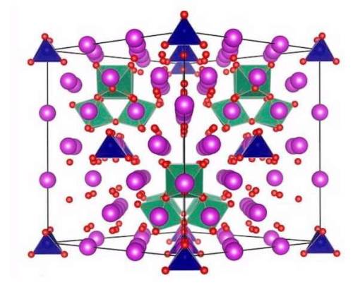
ANSTO has contributed to research led by the University of Sydney, involving doping transition metals in a polymorph of bismuth oxide in a search for more structural stability.
Cubic high-temperature polymorph of bismuth oxide, δ-Bi2O3, is the best known oxide ionic conductor but its narrow stability range (729—817 °C), which is close to its melting temperature of 817 °C precludes its practical use.
A large collaboration, led by Professor Chris Ling and Dr. Julia Wind (as part of her Ph.D.) from the University of Sydney involving researchers from ANSTO and two other universities, has achieved the design and understanding of the complex crystal structure and chemistry behind a commensurate structure within the fast-ion conducting stabilised bismuth oxide, co-doped with chromium and niobium, Bi23CrNb3O45.
The study was published in the Chemistry of Materials.
Post link
X-ray laser reveals how radiation damage arises
An international research team has used the X-ray laser European XFEL to gain new insights into how radiation damage occurs in biological tissue. The study reveals in detail how water molecules are broken apart by high-energy radiation, creating potentially hazardous radicals and electrically charged ions, which can go on to trigger harmful reactions in the organism. The team led by Maria Novella Piancastelli and Renaud Guillemin from the Sorbonne in Paris, Ludger Inhester from DESY and Till Jahnke from European XFEL is presenting its observations and analyses in the scientific journal Physical Review X.
Sincewater is present in every known living organism, the splitting of the water molecule H2O by radiation, called the photolysis of water, is often the starting point for radiation damage. “However, the chain of reactions that can be triggered in the body by high-energy radiation is still not fully understood,” explains Inhester. “For example, even just observing the formation of individual charged ions and reactive radicals in water when high-energy radiation is absorbed is already very difficult.”
To study this sequence of events, the researchers shot the intense pulses from the X-ray laser at the water vapor. Water molecules normally disintegrate on absorbing a single such high-energy X-ray photon. “Due to the particularly intense pulses from the X-ray laser, it was even possible to observe water molecules absorbing not just one, but two or even more X-ray photons before their debris flew apart,” Inhester reports. This gives the researchers a glimpse of what goes on inside the molecule after the first absorption of an X-ray photon.
Post link
Two-color X-rays give scientists 3-D view of the unknown
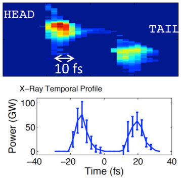
A high-resolution view of a sample or details of the first steps in ultra-fast processes are now available, thanks to researchers at SLAC National Accelerator Laboratory’s Linac Coherent Light Source (LCLS). They incorporated a series of advances to hit samples with a pair of precisely tuned X-ray laser pulses of different colors, or photon energies. The two pulses can arrive at the same time providing a detailed three-dimensional view of the sample’s structure. Delaying the beams by tens of femtoseconds (quadrillionths of a second) allows scientists to study very fast processes, such as the earliest steps of a chemical reaction.
For the first time, X-ray scientists have access and independent control of ultra-short two-color X-ray pulses with nearly the full peak power of the LCLS (above 50 GW). This new capability represents an improvement by an order of magnitude over what was possible before making it easier and faster for scientists to better dissect hard-to-study chemical and material science samples as well as map medically important proteins, such as signaling proteins of interest as pharmaceutical targets.
At the LCLS, researchers have demonstrated the generation of two-color X-ray pulses using twin electron bunches. In this technique, two electron bunches are generated by illuminating the injector photocathode with a train of two laser pulses separated in time by a few picoseconds. After being accelerated and compressed, the two electron bunches are spaced by tens of femtoseconds and have two distinct energies. When the high brightness bunched electron beams are delivered to a sequence of alternating magnets called an undulator, the two bunches emit two independent X-ray pulses of different photon energies.
Post link
Scientists observe nanowires as they grow
X-ray experiments reveal exact details of self-catalyzed growth for the first time
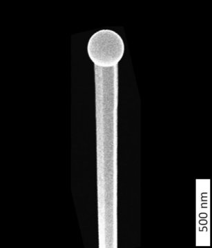
At DESY’s X-ray source PETRA III, scientists have followed the growth of tiny wires of gallium arsenide live. Their observations reveal exact details of the growth process responsible for the evolving shape and crystal structure of the crystalline nanowires. The findings also provide new approaches to tailoring nanowires with desired properties for specific applications. The scientists, headed by Philipp Schroth of the University of Siegen and the Karlsruhe Institute of Technology (KIT), present their findings in the journal Nano Letters. The semiconductor gallium arsenide (GaAs) is widely used, for instance in infrared remote controls, the high-frequency components of mobile phones and for converting electrical signals into light for fibre optical transmission, as well as in solar panels for deployment in spacecraft.
To fabricate the wires, the scientists employed a procedure known as the self-catalysed Vapour-Liquid-Solid (VLS) method, in which tiny droplets of liquid gallium are first deposited on a silicon crystal at a temperature of around 600 degrees Celsius. Beams of gallium atoms and arsenic molecules are then directed at the wafer, where they are adsorpted and dissolve in the gallium droplets. After some time, the crystalline nanowires begin to form below the droplets, whereby the droplets are gradually pushed upwards. In this process, the gallium droplets act as catalysts for the longitudinal growth of the wires. “Although this process is already quite well established, it has not been possible until now to specifically control the crystal structure of the nanowires produced by it. To achieve this, we first need to understand the details of how the wires grow,” emphasises co-author Ludwig Feigl from KIT.
Post link
Researchers gain a better understanding of the transformation of steel
Heating iron can alter its structure and is one of the methods for making various types of steel with different properties. That process is similar to the formation of frost flowers: one iron crystal structure transforms into another at a nucleus point, and the process expands further from that point. This type of nucleation in materials has the largest impact on their final properties, but is still the least understood in the field of metallurgy. For example, we still know little about how and where exactly this nucleation starts. Researchers at TU Delft have now shed new light on this subject in their publication ‘Preferential Nucleation during Polymorphic Transformations’ in Scientific Reports(Nature) of Wednesday 3 August.
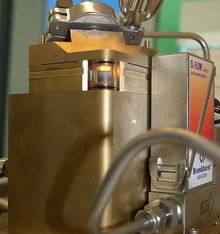
The researchers demonstrated this nucleation process live by heating an iron sample to 1,000 degrees in a specially produced furnace at the European Synchrotron Radiation Facility in Grenoble, and monitoring the transformation process using X-rays. In this way, they were able to identify the places and their properties that were the most likely starting points for the nucleation process of the transition from ferrite to austenite.
Post link
Images reveal battery materials’ chemical reactions in five dimensions – 3D space plus time and energy The chemical phase within the battery evolves as the charging time increases. The cut-away views reveal a change from anisotropic to isotropic phase boundary motion. Researchers at the U…
Novel X-ray lens sharpens view into the nano world
A team led by DESY scientists has designed, fabricated and successfully tested a novel X-ray lens that produces sharper and brighter images of the nano world. The lens employs an innovative concept to redirect X-rays over a wide range of angles, making a high convergence power. The larger the convergence the smaller the details a microscope can resolve, but as is well known it is difficult to bend X-rays by large enough angles. By fabricating a nano-structure that acts like an artificial crystal it was possible to mimic a high refracting power. Although the fabrication needed to be controlled at the atomic level — which is comparable to the wavelength of X-rays — the DESY scientists achieved this precision over an unprecedented area, making for a large working-distance lens and bright images. Together with the improved resolution these are key ingredients to make a super X-ray microscope. The team led by Dr. Saša Bajt from DESY presents the novel lens in the journal Scientific Reports (Nature Publishing Group).
“X-rays are used to study the nano world, as they are able to show much finer details than visible light and their penetrating power allows you to see inside objects,” explains Bajt. The size of the smallest details that can be resolved depends on the wavelength of the radiation used. X-rays have very short wavelengths of only about 1 to 0.01 nanometres (nm), compared to 400 to 800 nm for visible light. A nanometre is a millionth of a millimetre. The high penetration of X-rays is favoured for three-dimensional tomographic imaging of objects such as biological cells, computer chips, and the nanomaterials involved in energy conversion or storage. But this also means that the X-rays pass straight through conventional lenses without being bent or focussed. One possible method to focus X-rays is to merely graze them from the surface of a mirror to nudge them towards a new direction. But such X-ray mirrors are limited in their convergence power and must be mechanically polished to high precision, making them extremely expensive.
Post link
Liquid water at 170 degrees Celsius: X-ray laser reveals anomalous dynamics at ultra-fast heating
Using the X-ray laser European XFEL, a research team has investigated how water heats up under extreme conditions. In the process, the scientists were able to observe water that remained liquid even at temperatures of more than 170 degrees Celsius. The investigation revealed an anomalous dynamic behavior of water under these conditions. The results of the study, which are published in the Proceedings of the National Academy of Sciences(PNAS), are of fundamental importance for the planning and analysis of investigations of sensitive samples using X-ray lasers.
European XFEL, an international research facility, which extends from the DESY site in Hamburg to the neighboring town of Schenefeld in Schleswig-Holstein, is home to the most powerful X-ray laser in the world. It can generate up to 27 000 intense X-ray flashes per second. For their experiments, the researchers used series of 120 flashes each. The individual flashes were less than a millionth of a second apart (exactly 0.886 microseconds). The scientists sent these pulse trains into a thin, water-filled quartz glass tube and observed the reaction of the water.
“We asked ourselves how long and how strongly water can be heated in the X-ray laser and whether it still behaves like water,” explains lead author Felix Lehmkühler from DESY. “For example, does it still function as a coolant at high temperatures?” A detailed understanding of superheated water is also essential for a large number of investigations on heat-sensitive samples, such as polymers or biological samples.
Post link
Two X-ray techniques give a 3-D view of why catalysts used in gasoline production go bad
Merging two powerful 3-D X-ray techniques, a team of researchers from the Department of Energy’s SLAC National Accelerator Laboratory and Utrecht University in the Netherlands revealed new details of a process known as metal poisoning that clogs the pores of catalyst particles used in gasoline production, causing them to lose effectiveness.
The team combined their data to produce a video that shows the chemistry of this aging process and takes the viewer on a virtual flight through the pores of a catalyst particle. The results were published today in Nature Communications.
The particles, known as fluid catalytic cracking or FCC particles, are used in oil refineries to “crack” large molecules that are left after distillation of crude oil into smaller molecules, such as gasoline. Those oil molecules flow through the catalyst particles in tiny pores and passageways, which ensure accessibility to the active domains where chemical reactions can take place. But while the catalyst material is not consumed in the reaction and in theory could be recycled indefinitely, the pores clog up and the particles slowly lose effectiveness. Worldwide, about 400 reactor systems refine oil into gasoline, accounting for about 40 to 50 percent of today’s gasoline production, and each system requires 10 to 40 tons of fresh FCC catalysts daily.
Post link

X-ray of conjoined monkeys, Museum Vrolik.
During World War I, Marie Curie recognized that wounded soldiers were best served if operated upon as soon as possible. She saw a need for field radiological centers near the front lines to assist battlefield surgeons, including to obviate amputations when in fact limbs could be saved. After a quick study of radiology, anatomy, and automotive mechanics she procured X-ray equipment, vehicles, auxiliary generators, and developed mobile radiography units, which came to be popularly known as petites Curies (“Little Curies”). Read more here…
Post link

