#anatomy
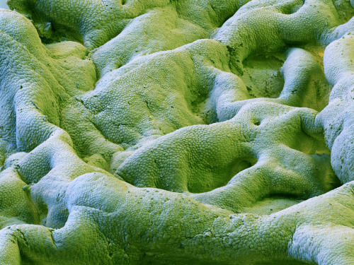
Image SS21025699 (Human Gallbladder SEM)
Scanning electron micrograph (SEM) of the surface of the gallbladder. Each of the small elevations on the wrinkled surface is an epithelial cell.
The gallbladder is a pear-shaped mucosal sac that serves as a reservoir for the bile fluid produced in the liver. Bile is released into the stomach after ingestion, where it plays an important role in the digestion of fats.
The bile emulsifies fats in partly digested food, thereby assisting their absorption. Bile consists primarily of water and bile salts, and also acts as a means of eliminating bilirubin, a product of hemoglobin metabolism, from the body.
Medicines and other “waste products” filtered out by the liver are also contained in the bile and are excreted in this way.
The gallbladder can be affected by gallstones, formed by material that cannot be dissolved, usually cholesterol or bilirubin. These may cause significant pain, particularly in the upper-right corner of the abdomen.
Magnification is 140x at a size of 12x12cm.
Image above © Eye of Science / Science Source
Post link
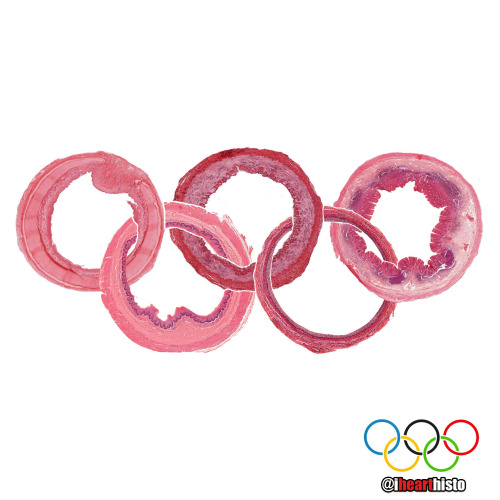
The Organ Rings
To commemorate the Olympic Games here are some slices through a few of the more tubular organs in your body!
Do you know what they are? Comment below!
Winner gets the gold medal!
[I’ll post the answers soon!]
Post link
Jurassic Pork
The pork tapeworm Taenia solium or a brachiosaurus?
i♡histo
This image shows the tapeworm T. solium, a parasite found in the bowels of humans who eat under-cooked pork.
You can see the proglottid segments of the worm’s neck/body and its terrifying scolex (head) complete with a hooked rostellum which it uses to attach itself onto your intestinal wall so that you don’t poop it out easily.
Fully grown worms can expand to around 2-3 meters in length and are composed of numerous segments each of which form their own reproductive unit.
The life cycle of the parasite begins when pigs ingest the T. solium eggs (from fecal contamination). The eggs develop into larvae inside the pig & then migrate through the intestinal wall to form cysts in their muscles & tissues.
Once slaughtered for meat, the larvae can be ingested by humans if the pork is eaten raw or under-cooked. Once in the human small intestine the larvae grow into large adult worms where they persist often unnoticed due to the lack of symptoms associated with their existence. The worms lay eggs into the intestines which are released during defecation thus continuing the life cycle.
“Life finds a way” - Dr. Ian Malcolm, Jurassic Park
A major complication of infection with T. solium is Cysticercosis. This is a parasitic tissue infection caused by the larval cysts of the tapeworm. These larval cysts infect brain, muscle, or other tissue, and are a cause of adult onset seizures. A person only gets cysticercosis by swallowing the eggs found in the feces of a person who has an intestinal tapeworm NOT from contracting a tapeworm by eating under-cooked pork itself. So people living in the same household with someone who has a tapeworm have a much higher risk of getting cysticercosis than people who don’t because they can be exposed to the eggs released in the stool.
Post link

Mi-fourth-sis
Dividing pyrotechnics!
Don’t forget to review the stages of mitosis while watching the fireworks this weekend!
It looks like this one is in metaphase!
Happy 4th of July!

❤️ Thanks Dad!
These are real spermatozoa.
Incidentally, based on head morphology, they are probably from a bull and not a human.
They were observed and photographed using phase contrast microscopy, a technique commonly used in semen analysis.
Note that the spermatozoa were realigned using photo editing software - spermatazoa are pretty cool cells but they do need a little help with their spelling sometimes.

SpongyBone SquarePants
Who lives in a bony medullary cavity?
Cancellous and yellow and porous is he.
Spongy bone!
Spongy bone is so named because its morphology (interconnected bony trabeculae/rods surrounding marrow spaces) resembles that of a sponge!
You can find spongy bone within flat bones (it forms the diplöe) and in long bones where it is located in the epiphyses (the ends of the bone) and diaphysis (the shaft of the bone).
Don’t be deceived by its name though. Spongy bone is not soft and spongy - it is still very strong, mature bone. It’s meshwork of trabeculae gives the inside of your bones strength and structure but at the same time, the space makes them more lightweight.
Spongy bone is always surrounded by a layer of compact bone (aka cortical bone).
Compact/cortical bone is different from spongy bone. It is named after the fact that it is more dense (i.e it isn’t made of rods and doesn’t have large spaces in it) and forms the outer aspect of a bone. If your bones were made entirely of compact bone than they would be much heavier and there would be no room for your bone marrow which is essential for making red and white blood cells.
Other names for spongy bone include ‘cancellous’ or 'trabecular’ bone.
Mother Earth
A biopsy of the mammary gland obtained during pregnancy.
Let’s celebrate our planet and home!
Happy Earth Day everyone!
i♡histo
Post link

Easter Bunny Blood Cell
This monocyte is declaring a warren germs!
And doesn’t carrot all about what he phagocytoses.
He’ll just hop right to it.
Original source of histology is unknown

Which came first?
A seasonal conundrum in some keratin debris within a benign lymphoepithelial cyst.
Happy Spring & Happy Easter everyone!
The image shows a swirl of keratin debris (the chicken) in a small epithelial cell nest (the egg). The salivary gland is packed full of lymphocytes (the many, many purple nuclei surrounding the epithelial nest) which are a type of white blood cell.
Salivary gland lymphoepithelial cyst like this are rare and benign. Once the cyst is removed surgically from the gland it rarely recurs.

Dinosmear
A rare sighting of the rawrsome Papanicolaous Rex!
The cells in this image are the squamous epithelial cells that line the region of the ectocervix the region of the hole (os) in the cervix where it protrudes into the vagina.
Doctors obtain these cells by scraping the cervix. The cells are then smeared onto a slide and stained with the Papanicolaou stain during the pap smear. Cytologists examine the cells for any signs of abnormal morphology that could be an indicator of cervical cancer or other pathology.
Image based on the original by @mik__e [Insta]
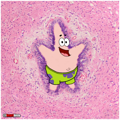
⭐ Patrick Star in a Sperm Tube
My favorite Patrick in honor of #stpaddysday☘️
Some say that Patrick Star and the male reproductive tract are fairly similar… but if you look closely there’s actually a vas deferens.
This image shows the tube through which spermatozoa travel during ejaculation. It is called the vas deferens (ductus deferens).
Histologically it is unique because of its unique pseudostratified epithelium (surrounding Patrick…check out the unique regular beaded appearance of the basal cells in this epithelium) and its triple-layered smooth muscle wall - so thick you can actually palpate it within the spermatic cord within the scrotum. Patrick is actually within the empty space of the lumen of the tube (where the sperm travels).
Spermatozoa are produced in the seminiferous tubules of the testes and travel to the epididymis where they complete the maturation process. During ejaculation, a peristaltic wave of strong muscular contractions propel the sperm from the epididymis and into the vas deferens. This wave of peristalsis continues along the triple-thick muscle wall of the vas deferens eventually pushing the sperm out into the urethra. From here they have enough momentum to travel along the length of the penis before exiting exit the tube at the external urethral meatus (the fancy anatomical name for the hole at the end of the penis).
But sperm are not the only stuff that is ejaculated… at the same time as the sperm is moving along this pathway, the smooth muscle surrounding a number of glands also contracts which pushes their secretions into the urethra too at the same moment that the sperm is moving through it. These include secretions from the seminal vesicles (seminal fluid), prostate gland (prostatic fluid) and bulbourethral glands.
The final concoction that is ejaculated is actually a combination of sperm and all of these extra sperm-nourishing secretions and collectively it is known as semen.
Which brings us back to where we started with this sea-man - Patrick Star!
#histology #science #pathology #pathologists #anatomy #autopsy #patrickstar #spongebob #vasdeferens #malereproductive #stpatricksday #menshealth #premed #biology #medicaleducation #meded #nurse #nursing #medschool #medstudent #medicine #medlab #vetscience #vetschool #ihearthisto
Post link
The Ro-lung Stones
The Stones’ ‘Hot Lips’ logo formed from a blood vessel filled with erythrocytes within a congested zone around a focal pneumonia of the lung.
During the first stages of focal/lobar pneumonia macrophages (the larger cells that are visible in the white alveolar space) respond to phagocytose (eat) any pathogens in the lung.
Additionally, the small blood vessels within the lung tissue begin to engorge (you can see all the erythrocytes in the dilated vessels not only in the large 'hot lips’ vessel but the smaller vessels in the interalveolar septa between adjacent alveoli). This is the congestion phase.
With this dilation of vessels come more white blood cells, you can see numerous neutrophils (they are the small cells that look like they have multiple lobes to their purple nuclei) have migrated out of the vessels (a process called extravasation) into the surround tissue and alveolar spaces. These cells are signs of an additional immune response and they help the macrophages destroy pathogens in the airway.
Image is by @beautiful_pathologist - check out her Instagram for more histology.
Post link

Happy Vagina Scalp Epididymis Liver!!
Here’s to a healthier, happier New Year for everyone.
Image shows:
2 - Blood vessel in vaginal mucosa
0 - Hair follicle in scalp
2 - Epididymis
1 - Hepatic portal vein branch in liver
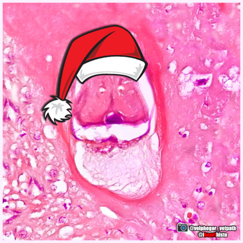
Santa Larvae
Hurry down my cecum tonight!
A nematode larva (Contracaecum) in the digestive tract of a seal. The eggs of this parasitic nematode use fish as an intermediate host before infecting piscivorous mammals, including humans who may forget to clean their holiday salmon!
This festive image was captured by veterinary pathologist @velphegor_vetpath via Instagram.
Happy holidays everyone!
Post link

A Charlie Brown Marrow:Happy Thanksgiving!
A developing leukocyte (white blood cell) observed in a sample of bone marrow obtained from the head of a femur
Blobfish-face
A coronal section through a developing jaw (when flipped upside down) looks like poor old blobfish!
The blobfish eyes are formed from Meckel’s cartilage. A component of the first pharyngeal arch that runs the length of the developing mandible. It degenerates as the fetus develops leaving only two small components on each side of the head. These ossify (become bone) to form the incus & malleus (ear ossicles) of the middle ear.
The blobfish nose is the developing tongue. It is composed of developing skeletal muscle fibers. Skeletal muscle forms from myoblasts that line up & fuse to form long myotubes. These will then synthesize actin/myosin which will allow them to contract & form the intrinsic skeletal muscle of the tongue.
The blobfish chin is formed by the developing maxilla. Two regions of tissue (the palatine shelves) grow together & fuse in the midline to form the posterior hard palate. You can see the midline suture forming & feel it in your own mouth by running your tongue along the roof of your mouth. Failure of these to fuse results in a variety of cleft lip and palate combinations.
The blobfish head is formed from a developing mandible. You can see small islands of bone forming within the mesenchymal tissue of the head. This type of bone development is called intramembranous ossification.
The blobfish is native to coastal waters off mainland Australia and Tasmania where it lives way deep down in the darkest depths of the ocean. Its gelatinous body is ideal for withstanding the pressure down there but when brought to the surface it looks like a sad melted pink crayon.
#histology #science #pathology #pathologists #anatomy #autopsy #blobfish #embryology #premed #biology #dentalschool #dentalstudent #dentistry #medicaleducation #meded #nurse #nursing #medschool #medstudent #medicine #medlab #vetscience #vetschool #vetstudent #histologia #histotech #histo #pathArt #ihearthisto
Post link
Caaaaarrrggghhh-diovascular Histology
Terrifying a medical student near you this Halloween!
The zombie’s lifeless eyes are arterioles.
His blood-filled mouth is a small vein.
All wrapped up in a connective tissue face.
Happy Halloween Histo fans!
Post link
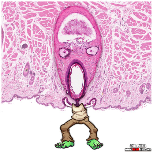
Zomb-hair
A very close-up scene from the hit TV series ‘The Walking Dreads’
This is actually a vibrissae hair follicle from the face of a (a whisker).
Vibrissae are sensory hairs that are different from regular body hairs in the fact that they are surrounded by a blood filled sinus (z’s brain) and are associated with sensory neurons that have a distinct and representative pattern in the somatosensory cortex of the mouse brain. The fact that they are well mapped in the brain illustrates their importance in everyday behavior and survival - they are involved in things like detecting, orienting and sensing length of surfaces/objects and tracking (e.g. finding gaps in a maze and answering the question 'can my head fit through here?’).
Note also the sebaceous glands (z’s eyes) which secrete a lipid rich secretion called sebum for maintaining hair and epidermis keratin (keratin is the wispy stuff lining the skin at the bottom of the image and forming z’s lil arms).
The hair in this follicle is absent as you can see z’s wailing mouth is completely empty (where you might expect to see the shaft of a hair).
The closest things us humans have to vibrissae are the thick long hairs in our nostrils which are fairly sensitive to tickles (and make your eyes water if plucked) but more importantly provide a first line of filtration to prevent big particles from being inhaled into your nasal cavity.
#histology #science #anatomy #pathology #autopsy #pathologists #dermatology #halloween #hair #skin #zombie #dentalstudent #dentalschool #histotech #medlab #premed #meded #nurse #nursing #medschool #medstudent #medicine #vetscience #vetschool #histotechnology #histologia #histo #pathArt #vetstudent #ihearthisto
Post link
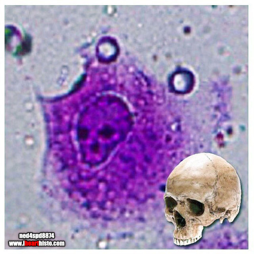
The Creepiest Fibroblast You Will Ever See
With a nucleus straight from the fiery depths of hell*
*Note: Fibroblasts do not come from hell. They are actually derived from embryonic mesoderm and the creepiest thing they do is secrete the collagen and ground substance of connective tissue. Not very creepy at all really when you think about it.
Histology is from the microscope of the ‘fiancee of ned4spd8834’
Post link

