#med school

Which came first?
A seasonal conundrum in some keratin debris within a benign lymphoepithelial cyst.
Happy Spring & Happy Easter everyone!
The image shows a swirl of keratin debris (the chicken) in a small epithelial cell nest (the egg). The salivary gland is packed full of lymphocytes (the many, many purple nuclei surrounding the epithelial nest) which are a type of white blood cell.
Salivary gland lymphoepithelial cyst like this are rare and benign. Once the cyst is removed surgically from the gland it rarely recurs.

Dinosmear
A rare sighting of the rawrsome Papanicolaous Rex!
The cells in this image are the squamous epithelial cells that line the region of the ectocervix the region of the hole (os) in the cervix where it protrudes into the vagina.
Doctors obtain these cells by scraping the cervix. The cells are then smeared onto a slide and stained with the Papanicolaou stain during the pap smear. Cytologists examine the cells for any signs of abnormal morphology that could be an indicator of cervical cancer or other pathology.
Image based on the original by @mik__e [Insta]
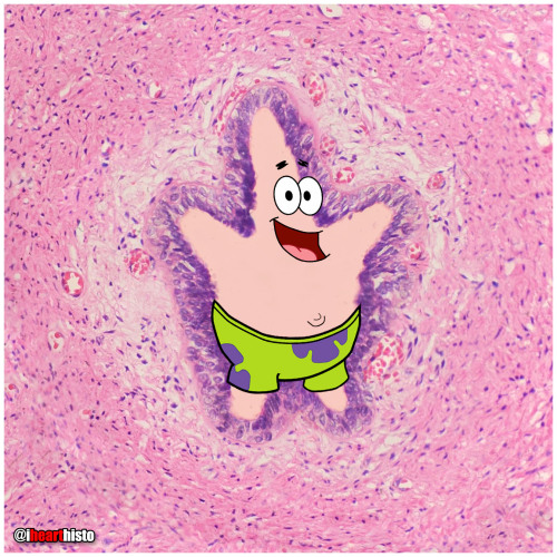
⭐ Patrick Star in a Sperm Tube
My favorite Patrick in honor of #stpaddysday☘️
Some say that Patrick Star and the male reproductive tract are fairly similar… but if you look closely there’s actually a vas deferens.
This image shows the tube through which spermatozoa travel during ejaculation. It is called the vas deferens (ductus deferens).
Histologically it is unique because of its unique pseudostratified epithelium (surrounding Patrick…check out the unique regular beaded appearance of the basal cells in this epithelium) and its triple-layered smooth muscle wall - so thick you can actually palpate it within the spermatic cord within the scrotum. Patrick is actually within the empty space of the lumen of the tube (where the sperm travels).
Spermatozoa are produced in the seminiferous tubules of the testes and travel to the epididymis where they complete the maturation process. During ejaculation, a peristaltic wave of strong muscular contractions propel the sperm from the epididymis and into the vas deferens. This wave of peristalsis continues along the triple-thick muscle wall of the vas deferens eventually pushing the sperm out into the urethra. From here they have enough momentum to travel along the length of the penis before exiting exit the tube at the external urethral meatus (the fancy anatomical name for the hole at the end of the penis).
But sperm are not the only stuff that is ejaculated… at the same time as the sperm is moving along this pathway, the smooth muscle surrounding a number of glands also contracts which pushes their secretions into the urethra too at the same moment that the sperm is moving through it. These include secretions from the seminal vesicles (seminal fluid), prostate gland (prostatic fluid) and bulbourethral glands.
The final concoction that is ejaculated is actually a combination of sperm and all of these extra sperm-nourishing secretions and collectively it is known as semen.
Which brings us back to where we started with this sea-man - Patrick Star!
#histology #science #pathology #pathologists #anatomy #autopsy #patrickstar #spongebob #vasdeferens #malereproductive #stpatricksday #menshealth #premed #biology #medicaleducation #meded #nurse #nursing #medschool #medstudent #medicine #medlab #vetscience #vetschool #ihearthisto
Post link
The Ro-lung Stones
The Stones’ ‘Hot Lips’ logo formed from a blood vessel filled with erythrocytes within a congested zone around a focal pneumonia of the lung.
During the first stages of focal/lobar pneumonia macrophages (the larger cells that are visible in the white alveolar space) respond to phagocytose (eat) any pathogens in the lung.
Additionally, the small blood vessels within the lung tissue begin to engorge (you can see all the erythrocytes in the dilated vessels not only in the large 'hot lips’ vessel but the smaller vessels in the interalveolar septa between adjacent alveoli). This is the congestion phase.
With this dilation of vessels come more white blood cells, you can see numerous neutrophils (they are the small cells that look like they have multiple lobes to their purple nuclei) have migrated out of the vessels (a process called extravasation) into the surround tissue and alveolar spaces. These cells are signs of an additional immune response and they help the macrophages destroy pathogens in the airway.
Image is by @beautiful_pathologist - check out her Instagram for more histology.
Post link

Happy Vagina Scalp Epididymis Liver!!
Here’s to a healthier, happier New Year for everyone.
Image shows:
2 - Blood vessel in vaginal mucosa
0 - Hair follicle in scalp
2 - Epididymis
1 - Hepatic portal vein branch in liver
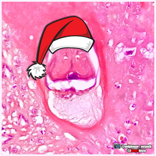
Santa Larvae
Hurry down my cecum tonight!
A nematode larva (Contracaecum) in the digestive tract of a seal. The eggs of this parasitic nematode use fish as an intermediate host before infecting piscivorous mammals, including humans who may forget to clean their holiday salmon!
This festive image was captured by veterinary pathologist @velphegor_vetpath via Instagram.
Happy holidays everyone!
Post link

A Charlie Brown Marrow:Happy Thanksgiving!
A developing leukocyte (white blood cell) observed in a sample of bone marrow obtained from the head of a femur
Caaaaarrrggghhh-diovascular Histology
Terrifying a medical student near you this Halloween!
The zombie’s lifeless eyes are arterioles.
His blood-filled mouth is a small vein.
All wrapped up in a connective tissue face.
Happy Halloween Histo fans!
Post link
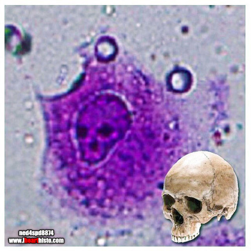
The Creepiest Fibroblast You Will Ever See
With a nucleus straight from the fiery depths of hell*
*Note: Fibroblasts do not come from hell. They are actually derived from embryonic mesoderm and the creepiest thing they do is secrete the collagen and ground substance of connective tissue. Not very creepy at all really when you think about it.
Histology is from the microscope of the ‘fiancee of ned4spd8834’
Post link
It’s amazing how a simple thing like grabbing an extra coffee for your consultant who is working the night shift goes a long way.
I heard on a podcast once that the way to be kind is to treat kindness like a muscle that you have to train.
It’s so easy to throw yourself a pity party but it leads you nowhere (I’m guilty of this). It feels way better to step outside of yourself, consider other people’s circumstances and be kind. I hope I can always remember to be kind.
Post link
“It’s nothing personal.”
Something we hear often after being yelled at for something entirely out of our control. I’ve been seeing some news about physician abuse. Whether verbal or emotional or physical, it happens. And it sucks.
But here’s the thing. A lot of times there are laws to protect healthcare workers from abusive patients. But what happens when the abuse comes from fellow colleagues? Senior doctors who feel entitled to demean anyone with a lesser job title? What laws protect us as students from being made to feel inadequate every time we don’t know the right answer? Why are we promoting this idea that scolding/yelling/being mean is the way to make sure we learn?
We are promoting a whole different type of abuse and making it seem like it’s okay because hey, it’s nothing personal right?
I hope we can find it in us to be kind to everyone around us this week..
Follow on instagram: https://www.instagram.com/mindofamedstudent/
Post link

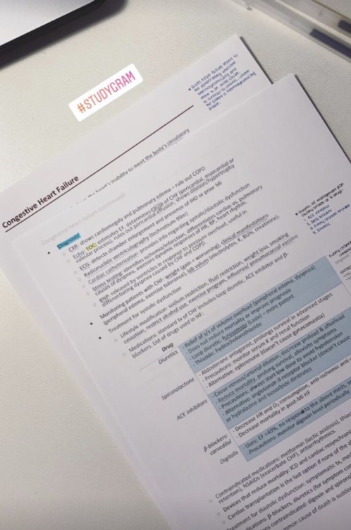
I love writing notes. I actually enjoy the process of summarizing pages of textbook information and ending up with a concise few pages that I can refer to for studying. One of the questions I’ve seen in my inbox quite a few times is whether I prefer to write or type my notes. It’s interesting to me because I actually recently switched back to mainly typing notes after writing for such a long time.
Because I’ve done and still do both, I figured there must be a reason I can’t choose one over the other. To solve this dilemma, I decided to make a pro/con list for each and share it as a way of answering these questions.
Handwriting
Pros
- Engaging my senses in the learning process by writing increases the chances that I’ll remember what I’m noting down.
- It’s faster for drawing things out - specifically processes and flowcharts.
Cons
- It takes me more time, specially because I try to be very neat.
Typing
Pros
- It almost always takes me less time.
- It’s easier to back up/share notes this way.
- It’s a more convenient way of adding in pictures.
- It’s easier to edit later on.
Cons
- Using a tablet/laptop provides easier access to distraction.
- Staring at the screen for too long gives me a headache.
There’s nothing like the feel of pen to paper… But I found that eventually, I still need to spend time revising by going over my notes whether I’ve handwritten or typed them. So it seems like a better use of my time to finish my summaries faster in order to be able to start “actually studying”. So why not do the thing that saves time?
My biggest reason for switching back though has been the following. In my last two years of medical school, I’m teaching myself a lot of theoretical information from books. But I also go to the hospital and see those cases, so I’m constantly learning new information and making new links that I didn’t make before (as in, when I was learning the information from a textbook). Because of this, I end up having to annotate notes a lot. At first it didn’t seem like an issue. But eventually when my notes were all over the place, it was starting to become disorganized and I didn’t like that. But that’s just me!
And as always.. Disclaimer: I do things this way because of my own priorities. By no means am I claiming everybody should do the same or that it’s perfect. I’m just providing ideas and I hope they help :)
Ps. I’m now on instagram! Where I’m trying to post daily stories and have a visual-based companion to this blog. Follow me here https://www.instagram.com/mindofamedstudent/
I’m on Instagram!
Follow me there for daily updates on my story :)
https://www.instagram.com/mindofamedstudent
Post link
Zinc supplementation may exacerbate rheumatoid arthritis (RA), new laboratory data suggest.
In monocytes from rheumatoid arthritis patients, plasma zinc concentrations and Zip8 expression were increased, and Zip8 expression correlated with more severe disease. Thus, inhibiting zinc influx into monocytes and macrophages could prevent excessive inflammatory responses that occur in diseases such as rheumatoid arthritis – the researchers concluded.
A team of doctors and researchers working at Erasmus Hospital in Belgium has successfully treated an adult woman infected with a drug-resistant bacteria using a combination of bacteriophage therapy and antibiotics. In their paper published in the journal Nature Communications, the group describes the reasons for the use of the treatment and the ways it might be used in other cases.
Bacteriophages are viruses that infect and kill bacteria. Research involving their use in human patients has been ongoing for several decades, but they are still not used to treat patients. In this new effort, the researchers were presented with a unique opportunity not only to treat a patient in need of help, but to learn more about the possible use of viruses to treat patients infected with bacteria that have become resistant to conventional antibiotics.
Bacteria may Demonstrate any of Five General Mechanisms of Antibiotic Resistance:
1. Lack of entry; Decreased cell permeability.
2. Greater exit; Active efflux.
3. Enzymatic inactivation of the antibiotic.
4. Altered target; Modification of drug receptor site.
5. Synthesis of resistant metabolic pathway.
More than HIV, more than malaria. The death toll worldwide from bacterial antimicrobial resistance (AMR) in 2019 exceeded 1.2 million people, according to a new study.
In terms of preventable deaths, 1.27 million people could have been saved if drug-resistant infections were replaced with infections susceptible to current antibiotics. Furthermore, 4.95 million fewer people would have died if drug-resistant infections were replaced by no infections, researchers estimated.
A new type of diagnostic blood test has been shown to accurately detect cancer in patients with non-specific symptoms, such as unexplained weight loss and fatigue, as well as differentiating between patients with localized and metastatic disease.
This makes it the first blood-based cancer test to determine the metastatic status of a cancer without prior knowledge of the primary cancer type.
In a study published this week in the journal Clinical Cancer Research, researchers from the University of Oxford analyzed blood samples from 300 patients with non-specific but concerning symptoms of cancer using a technique called nuclear magnetic resonance (NMR) metabolomics.
Unlike conventional blood-based tests for cancer, which look for genetic material from tumors, the NMR-based technique uses magnetic field and radio waves to analyze levels of metabolites in the blood as biomarkers to distinguish between different cancer states.

Answer and Explanation:


